


This is a rare disease in the UK with cases found here since 2012. It is a seasonal disease with the majority of cases being identified between November and May. The general advice is that if your dog develops unexplained skin lesions especially on its legs, although other parts of its body can be affected, please check it out with your vet. It’s important that your dog is treated early as once the kidneys are affected much more intensive treatment is required which may not be successful.
There are a number of websites with pages dedicated to Alabama Rot, the latest being hosted by the vets that have been closely connected with the diagnosis and treatment of the disease since it was first diagnosed in the UK.
https://www.alabama-rot.co.uk/
https://www.medivet.co.uk/pet-care/pet-advice/alabama-rot-map/
https://www.rvc.ac.uk/small-animal-vet/news/alabama-rot-in-the-uk-frequently-asked-questions
Update on Bloat July 2022
Professor Ed Hall, our Breed Health Co- ordinator, attended the recent Large and Giant Breed Working Group meeting and one of the speakers was Professor Mark Dunning who gave a presentation on bloat. He is working with The Deerhound Club investigating the factors influencing bloating and the development and outcome of GDV in Deerhounds in the UK. Below are the minutes concerning his presentation and reproduced with kind permission from the Kennel Club.
Gastric Dilatation Volvulus (GDV)/ Bloat in Large and Giant Breeds
Prof Dunning gave a presentation to the group regarding current knowledge surrounding GDV.
He began by differentiating bloat and GDV, explaining that GDV, an acute life-threatening disease, results from serious bloat (or GD). The mechanisms of GDV were described, with the stomach turning on to its left side, which results in the oesophagus becoming twisted, preventing any air escaping out of the stomach and escalating the bloating. This causes extreme stretching of the tissues and displacement of the spleen and other organs. The following YouTube video was shared to show the movement: Patterson Veterinary DIA Client Education Video- Gastric Dilatation-Volvulus (GDV)- Bloat – YouTube
Torsion is incredibly serious as the movement of the stomach causes a stretching of the gastric wall, which prevents blood circulation and oxygen reaching tissues properly, leading to necrosis (cell death), clots, haemorrhage and possible peritonitis/ septicaemia. This leads to a wider obstruction of blood flow across the body of an affected individual, causing an elevated heart rhythm, whole-body inflammation and poorer chance of survival.
There is no simple solution for every dog but there are multiple factors that lead to an outcome, and therefore by understanding the general indicators we can better mediate and treat affected individuals. Risk factors include both intrinsic (within the body) and extrinsic (i.e. environmental) factors. Extrinsic factors include diet, frequency of feeding, type of food, elevated/ low-level feeding, exercise, history of or ongoing chronic gastrointestinal disease, and aerophagia (gulping air). Intrinsic factors are more difficult to change as they are inherent to the individual, these factors include the following: breed, the genetics of the immune system (i.e. whether it takes form in an autoimmune state and begins attacking tissues/ organs within the body, such as within the gut), body size, being deep-chested, gastric volume and position, how laxly the stomach is attached to the abdomen, being able to release gas, familial history of GDV, and temperament and stress.
MD noted that as such GDV is incredibly complex, and cannot be avoided by merely reducing one factor, but by reducing the risk of all the above where possible it is more likely to minimise overall risk.
With respect to research, MD noted there is a trend investigating the gut microbiome (bacteria populations) in affected dogs and underlying genetic components, pointing towards difficult inherent factors. There have been certain differences in gut microbiome found between GDV affected and unaffected dogs in recent years, which are being more easily identified thanks to advancements in technology and analysis. Future research and establishing the association between gastrointestinal flair-ups with GDV could allow for better prophylactic (preventative) treatments. The importance of educating owners in normalising abnormal underlying disease (e.g. ongoing loose stools) was also highlighted as being important due to the established links between such disease and GDV risk. This research indicates an element of immune dysfunction and that further genetic research is necessary to establish whether any genes that maintain the immune system also influence GDV. Several recent papers have begun to identify a number of genes associated with GDV affected dogs.
A limitation of research is that many breeds affected have very numerically small populations, and therefore the disease has not been well described in some of these breeds. With this, much of MD’s research is to work with the breeds and BHCs to access these data, which would otherwise be lost through veterinary practice/ hospital datasets.
MD then gave an overview on outcomes in affected dogs and how mortality rates have changed overtime post-surgery. Certain procedures will alter the likelihood of outcome, with removal of organs (i.e. parts of the stomach/ spleen) further increasing the risk of mortality. Outside of surgery other factors that can impact prognosis include, how quickly a dog is presented to the vets, and whether the dog is showing signs of critical inflammation/ hypotension/ sepsis/ peritonitis/ necrosis and splenic trauma. A further factor identified has been arrhythmia which can lead to an increased risk of mortality. Echocardiogram is needed to identify any dangerous or unstable arrhythmias, particularly any abnormalities where the heart is attempting to contract during its relaxed phase – these arrhythmias will require intervention to prevent fatality.
Sadly, MD reported that studies have found 50% of dogs presented with GDV are euthanised on arrival, without undergoing any further treatment. There are a number of factors which lead towards this, primarily being cost and/ or being uninsured, and time of day (i.e. cases presented overnight are less likely to survived). Longer surgeries have also been suggested to negatively influence a dog’s likelihood of survival, however MD did note that there will be variability in a surgeon’s/ support team’s skills which makes data difficult to interpret.
Biomarkers available that help predict a dog’s outcome post-surgery include CRP, lactate, cPLI, and procalcitonin which give indication for the amount of inflammation and circulatory distress occurring in the body. Having the ability to measure these will allow the vet team to further assess a dog’s clinical status and gives strategic measurements, which can be taken into account for a dog’s recovery, and give a better prediction of a dog’s likelihood to survive.
MD then walked the group through the process from development of symptoms to presentation at veterinary level. Diagnosis is needed to differentiate between bloating and GDV. Primary clinical signs included non-productive and continuous retching/ vomiting, a progressive abdominal swelling, pain, depression, pacing, and being in shock. MD noted that having even a small inkling of something being wrong is enough to warrant taking a dog to the vet, as it has been proven that early action is key to survival.
Surgeons on presentation will be looking to restore circulation and oxygen to tissues, decompressing the stomach via a stomach tube, determining whether the disease is GD or GDV through imaging (X-ray or CT scan), surgical correction if needed, and further prophylaxis to prevent any future episodes. If lavage via a stomach tube is not effective, letting the air out through a needle in the abdomen is also an effective method. During surgery a surgeon will untwist the stomach, then look at whether any stomach/ spleen tissue needs removing, and then fix the stomach via gastropexy to prevent any further GDV episodes.
Prophylactic gastropexy is becoming more widely used and can be undertaken at the time of neutering as part of a routine procedure. MD did note that incisional gastropexies appear to be less effective than other prophylactic gastropexies, however further work is needed here to tease apart efficacy. MD did go on to note that any form of pexy is going to give a better chance than no prophylaxis. Studies that have monitored dogs that have had appropriate gastropexies have found that none of these have gone on to develop GDV. It was noted that gastropexy should not be undertaken on dogs that have not yet reached maturity.
MD went on to discuss ‘low-hanging fruit’ (e.g. epidemiology) and complex investigations (e.g. genetics) research and management, and that both of these approaches should be made in tandem to manage disease risk. It was noted that as this is not a simple genetic disorder and cannot be selected against to be completely eradicated, but can be taken into account to produce less-predisposed progeny, and continuing to manage extrinsic factors will help to minimise risk.
MD’s research suggest that different breeds have different factors contributing towards disease risk; for example for Deerhounds a recent stressful event has been thought to contribute more heavily to disease, in comparison to Greyhounds where having a history of abdominal surgery contributes further. Identifying such subtle signs and pointers will allow owners to manage their animals with this in mind, and determine whether and how quickly they need to get their dog to the vets. MD’s student is working on analysis for a number of other breeds, where it is hoped that other findings can be established.
The importance of improving communications between owners, breeders and vets was stressed, to allow a more complimentary approach to breed care. As such, MD was keen to work with the BHCs to improve breed-specific understanding. Any breeds that would like to collaborate with MD’s project should email Hannah who will put them in touch with MD’s team. Many breeds have worked on their own surveys which have been very useful in gauging prevalence and identifying initial factors involved in their breed’s risk, however it was mentioned that to allow a consistent and universal approach it would be beneficial to make use of MD’s team and resourcing to further expand on this work.
The floor was then opened up to the breed representatives for any questions or comments.
A query was raised with respect to raised feeding, with MD noting that the picture is mixed, and should be tailored to an individual dog. The most important factor to consider is whether a dog is aerophagic or not therefore owners should observe whether the dog gulps food. To manage this factor owners should aim to slow down rate of feeding and evaluate whether raised/ low-level feeding improves this.
The picture surrounding diet is still complex, but increasing the amount of time that the body takes to digest food is thought to have an impact. The group queried what the current knowledge is in respect to raw versus kibble and risk. MD noted that raw is a difficult factor to pick apart as there are a number of different forms of raw, alike to kibble. At this stage anecdotally dogs that move onto raw have less gastrointestinal disease, however long-term studies are needed to follow dogs overtime to try to identify influence. Underlying intestinal health certainly appears to have an influence on development of GDV, but further work is needed in this area. There are also subtleties in diet, with many dogs fed extra supplements/ bits of raw, kibble etc. so obtaining a study group with set and consistent feeding patterns will be a difficult objective to meet.
“Simple” genetic gene tests have been said to be available, however the efficacy of these have not been published in the peer-reviewed literature, and given that the condition is thought to be a complex disease with multiple interplaying genes, it is unlikely that these tests are, at present, good predictors of disease.
Many of the BHCs reported in their own experience that stress had seemed to be a contributing factor, and the importance of knowing your own animal and improving understanding owner awareness and husbandry.
The group thanked MD for his time and willingness to join the group to share his experience.
_________________________________________________________________________________________
About Bloat
Bloat is a very serious health risk for many dogs, but especially large and giant breeds. Unfortunately the Irish Setter is one of the breeds that is particularly prone, and it is really important that owners are aware of and can recognise the signs so they can contact their vet immediately, day or night, if they think their Setter is blowing. Getting your Setter to the vet immediately is crucial as time is absolutely vital; don’t wait to “see what happens” and certainly don’t wait to see if your dog is better in the morning. Bloat is an emergency and all vets are aware of the importance of seeing the dog immediately.
This is a complex disease which is likely to be the result of environmental influences including diet and stress as well as familial susceptability. Whilst it is not clear whether it is truly inherited, or whether it is a reflection of inherited conformational characteristics, it does mean that the chances of your puppy getting bloat increase if there have been other cases of bloat in the family.
What names is bloat known by?
- Bloat
- Dilatation-Volvulus
- Distension
- Gastric Dilatation
- Gastric Torsion
- Gastric Volvulus
- GDV
- Torsion
- Tympani
These are all names that may be used to describe bloat and are often used interchangeably by owners as they are all stages of Gastric Dilatation-Volvulus (GDV)
What is Bloat?
- Bloat is an unusual accumulation of gas and fluids in the stomach which is not passed normally through belching or flatulence and which causes abnormal swelling.
- The gas that accumulates is largely swallowed air, and does not arise by fermentation in the stomach. The stomach becomes like a drum (tympani).
- A dilatation is where the stomach is distended but is not twisted.
- Eventually the stomach will not only just dilate but also rotate fully on its long axis, causing a torsion/volvulus.
A bloated stomach affects several other organs in the abdomen by putting pressure on them and by affecting the veins which means blood cannot return to the heart as it should. The spleen may also become twisted. As the stomach gets bigger it puts pressure on the chest cavity which makes it difficult for the dog to breathe. If the stomach twists it can totally or partially block the exit to and from the stomach trapping gas, food and water in the stomach. The stomach’s own blood supply can be comprised leading to death of its wall, rupture and peritonitis. This combination can quickly lead to death as organ failure, low blood pressure and shock all set in.
Symptoms
Not all dogs get all the symptoms so don’t wait to see them all:
Phase 1:
The stomach is dilating but may not have twisted.
- Not acting as normal
- Restlessness and anxiety
- May ask to go outside in the middle of the night
- Swelling of the stomach- feels like a drum and may resonate when tapped gently
- Excessive salivation
- Pacing
- Stretching
- Looking at abdomen
- Whining
- Unproductive retching: attempts to vomit but not bringing up food; sometimes a white, frothy saliva is brought up
Phase 2:
The stomach has twisted and shock is starting to set in
- Abdominal pain
- Very restless
- Whining and groaning
- Pacing
- Unable to settle
- Stretching
- Looking at the abdomen
- Abdomen is enlarged and tight
- Difficulty in breathing
- Panting
- May stand with front legs apart and head down
- Trying to vomit more often
- Heart rate increased to 80 – 120 beats per minute
- Dark red gums
Phase 3:
Shock has developed and death is imminent
- Shallow breathing
- White or blue gums
- Weak pulse
- Abdomen is very enlarged
- Heart rate over 120 beats per minute
- Collapse
Measures thought to reduce the risk of bloat.
- Feeding two or three smaller meals a day rather than one large one
- Avoiding exercise for a couple of hours before and after feeding
- Limiting the amount of water available immediately before and after eating
- Feeding a good quality diet
- Not feeding a meal that swells in contact with water
- If you are changing diet then doing it gradually over a period of a few days
- Making meal times as stress free as possible
- Making sure your Setter is not underweight
- If you have more than one dog and there is a race to finish eating then it is best to separate them
It used to be thought that feeding your dog from a raised bowl helped to prevent bloat but more recent research shows this is not the case.
Treatment
Urgent veterinary attention should be sought if you think your Setter is bloated.
Emergency treatment will comprise intravenous fluids to compensate for shock and decompression of the stomach by a stomach tube. Surgery to correct any torsion will be performed as soon as the dog is stable.
General Information
- Dogs that bloat are generally over 2 years old and the chance increases by the time they are about 4 but this is not always the case. Puppies have been known to bloat and, occasionally, dogs over 10 will bloat.
- Larger deeper chested dogs seem to be most at risk.
- There may be a history of digestive upsets, but this in not always the case.
- Having a first degree relative (i.e. parent, sibling) with a history of bloat seems to increase the chances of bloat.
- There may be a familial association with other dogs who bloat but this is not always the case.
- Stress is a known factor and “happy dogs” are considered to be less at risk.
Prognosis and prevention of recurrence
Bloat is a serious condition, with a mortality rate of approximately 30% even with prompt veterinary treatment, although the prognosis is worsened if treatment is delayed.
Dogs that survive an episode of bloat are at increased risk of repeat episodes. The risk can be significantly reduced by performance of a surgical procedure, called a gastropexy, that fixes the stomach’s position and prevents it from twisting, although it will not prevent further episodes of dilatation. This procedure is performed either at the time of the first surgery, or at a later date if a patient is treated with fluids and decompression initially. It is important that you request that your vet perform this surgery which may be performed.
You may be given the option of laparoscopic gastropexy. Commonly called keyhole surgery it is minimally invasive, faster and has better healing results.
The x rays below, courtesy of the vet who took them, shows the before and after scenario of an Irish setter which bloated. That on the left clearly shows the distended stomach which is the large black mass to the right on the x ray. That on the right was taken after the stomach was decompressed. In this case the stomach hadn’t twisted and a laparoscopic gastropexy was carried out a few days later.

Bloat surveys and research from Purdue University
Raghavan, M.; Glickman, N.W.; Glickman, L.T. The effect of ingredients in dry dog foods on the risk of gastric dilatation-volvulus in dogs. Journal of the American Animal Hospital Association, 42: 28-36, January/February 2006.
Glickman, L., Glickman, N., et al. Non-dietary risk factors for gastric dilatation-volvulus in 11 large and giant breed dogs. Journal of the American Veterinary Medical Association, 217(10):1492-1499, 2000.
Glickman, L.T., Glickman, N.W., Schellenberg, D.B., Raghavan, M., Lee, T.L. Incidence of and breed-related risk factors for gastric dilatation-volvulus in dogs. Journal of the American Veterinary Medical Association, 216(1):40-45, 2000
Schellenberg, D., Yi, Q., Glickman, N.W., Glickman, L.T. Influence of thoracic conformation and genetics on the risk of gastric dilatation-volvulus in Irish setters. Journal of the American Animal Hospital Association, 34(1):64-73, 1998.
Glickman, L.T.; Lantz, G.C.; Schellenberg, D.B; Glickman, N.W. A prospective study of survival and recurrence following the acute gastric dilatation-volvulus syndrome in 136 dogs. Journal of the American Animal Hospital Association, 34(3):253-9, 1998
Schaible, R.H.; Ziech, J.; Glickman, N.W.; Schellenberg, D.; Yi, Q.; Glickman, L.T. Predisposition to gastric dilatation-volvulus in relation to genetics of thoracic conformation in Irish Setters. Journal of the American Animal Hospital Association, 33(5):379-83, 1997
Glickman, L.T.; Glickman, N.W.; Perez, C.M.; Schellenberg, D.S.; Lantz, G.C. Analysis of risk factors for gastric dilatation and dilatation-volvulus in dogs. Journal of Veterinary Medical Association 204(9):1465-71, 1994
Raghavan, M.; Glickman, N.W.; Glickman, L.T. The effect of ingredients in dry dog foods on the risk of gastric dilatation-volvulus in dogs. Journal of the American Animal Hospital Association, 42: 28-36, January/February 2006.Bloat surveys and research from Purdue University
Glickman, L., Glickman, N., et al. Non-dietary risk factors for gastric dilatation-volvulus in 11 large and giant breed dogs. Journal of the American Veterinary Medical Association, 217(10):1492-1499, 2000.
Glickman, L.T., Glickman, N.W., Schellenberg, D.B., Raghavan, M., Lee, T.L. Incidence of and breed-related risk factors for gastric dilatation-volvulus in dogs. Journal of the American Veterinary Medical Association, 216(1):40-45, 2000
Schellenberg, D., Yi, Q., Glickman, N.W., Glickman, L.T. Influence of thoracic conformation and genetics on the risk of gastric dilatation-volvulus in Irish setters. Journal of the American Animal Hospital Association, 34(1):64-73, 1998.
Glickman, L.T.; Lantz, G.C.; Schellenberg, D.B; Glickman, N.W. A prospective study of survival and recurrence following the acute gastric dilatation-volvulus syndrome in 136 dogs. Journal of the American Animal Hospital Association, 34(3):253-9, 1998
Schaible, R.H.; Ziech, J.; Glickman, N.W.; Schellenberg, D.; Yi, Q.; Glickman, L.T. Predisposition to gastric dilatation-volvulus in relation to genetics of thoracic conformation in Irish Setters. Journal of the American Animal Hospital Association, 33(5):379-83, 1997
Glickman, L.T.; Glickman, N.W.; Perez, C.M.; Schellenberg, D.S.; Lantz, G.C. Analysis of risk factors for gastric dilatation and dilatation-volvulus in dogs. Journal of Veterinary Medical Association 204(9):1465-71, 1994
Breed
For several years now the health of purebred dogs has been in the spotlight and the UK Kennel Club requested that each recognised breed appoint a health committee to examine all health issues in their breed. Our committee was set up with health representatives from the eight Irish Setter breed clubs in the United Kingdom and we have an independent chairman Prof. Edward J Hall MA, Vet MB, PhD, Dip ECVIM-CA, MRCVS.
Ed Hall is a professor of Small Animal Internal Medicine at the University of Bristol Veterinary School, where he is Head of the Division of Companion Animal Studies. A Cambridge graduate he undertook clinical and research training in Philadelphia and Liverpool, and is a diplomate of the ECVIM -CA. In his PhD he investigated glutensensitive enteropathy in Irish setters with great assistance from Mrs Jean Quinn and other breeders. This is where his passion for the breed developed.
He is currently a past president of the British Small Animal Veterinary Association and has written over 70 scientific papers and numerous book chapters chapters on canine small intestinal diseases.
He has clinical and research interests in small animal gastroenterology, in particular inflammatory bowel disease, and sees referral patients with GI problems. He gave evidence to the APGAW and Bateson enquiries into pedigree dog breeding. He owns an Irish Setter Fin.
Health topics of interest and importance to Irish Setter breeders and owners are discussed at the meetings and one of the first actions of the group was to organise an online survey for all Irish setters owners to” obtain a snapshot of the state of Irish setter health with respect to known and suspected inherited disease. They also wish to identify those conditions that setter owners believe have the most serious impact on the health and welfare of their dogs, so that future initiatives can be targeted at the most important conditions.” this information has been analysed and presented to the committee and Ed Hall has written a report.
The committee also supported Cathryn Mellersh from the AHT in the genetic investigation of bloat in the Irish Setter and believe it is the first investigation to consider the probable genetic component.
If you have concerns about any aspect of the health of this breed, or your Irish Setter, which you consider should be brought to the attention of the Health Committee or require more information on any health matter, please feel free to contact any breed club health representative or SEISC Health Representative Meg Webb megwebb1@aol.com. You do not need to be a member of any club.
Breed Health Coordinator:
Emeritus Professor Ed Hall, Department of Clinical Veterinary Science, University of Bristol
Club Health Representatives:
Belfast and District Irish Setter Club: Karen McKelvey
Irish Setter Association, England: Gemma Gisby
Irish Setter Breeders Club: Jo-Anne Parsons
Irish Setter Club of Scotland: Chloe Green and Jenna Sturrock
Irish Setter Club of Wales: Graham Hart
Midland Irish Setter Society: Stephen Wood
North East of England Irish Setter Club: Sally Mohan
South of England Irish Setter Club: Meg Webb
The Kennel Club (KC) and the British Veterinary Association (BVA) run health schemes for hip dysplasia, elbow dysplasia and eye diseases which provide scientifically based expert opinion on these inherited conditions. The KC/BVA screening programmes help conscientious breeders to identify dogs that are clinically free of such diseases so that the best possible choices for breeding programmes may be made. All results are recorded on the Kennel Club database and published in the Kennel Club Breed Records Supplement.
Hip Dysplasia Scheme
It is only necessary to hip score once for each dog in its lifetime but only after it is a year old. An X-ray is required for this which must be submitted by the owner’s vet to a KC/BVA panel which reads it and gives individual scores for each hip. The maximum score for each hip is 53 giving a maximum total of 106 and the lower the score the better the hips. Each breed has a Breed Mean Score (BMS), this being the average of the total hip scores. The KC encourages breeders to only breed from dogs which have a score lower than the BMS; the BMS for Irish Setters is currently 11.
Details of the Hip dysplasia scheme can be found at:
https://www.bva.co.uk/media/4891/hip-breed-specific-statistics-2022.pdf
Elbow Dysplasia Scheme
It is only necessary to elbow grade once for each dog in its lifetime but only after it is a year old. Irish Setters are not known for elbow problems and this test is rarely carried out by breeders. An X ray is required which must be submitted by the owner’s vet to a KC/BVA panel which reads it and gives individual scores ranging from 0-3.
Eye Scheme
It is recommended that eye examinations should, in general, be conducted annually. Dogs are certified either affected or unaffected for the eye conditions that are known to be inherited in the breed at the time of examination. Re-examination is important as some inherited eye conditions have a later onset.
DNA Screening Schemes
DNA tests give precise information as to the genetic status of dogs tested with regards to specific diseases. Detailed information as to how these tests work can be found in: DNA Testing and Autosomal Recessive Genes. Irish Setters breeders have 3 DNA tests which are available and each of the three diseases below is described as an autosomal-recessive condition. This means that a dog must inherit two copies of an abnormal gene (one from its mother and one from its father) before its health is affected. A dog that inherits only one copy of the abnormal gene (from its mother or its father) will have no signs of the disease, but will be a carrier and may pass the gene on to any offspring.
PRA rcd1 (Progressive Retinal Atrophy) rod cone dysplasia 1
See our page: PRA Blindness for details of rcd1.
CLAD (Canine Leucocyte Adhesion Deficiency)
See our page: Canine Leucocyte Adhesion Deficiency (CLAD) for details
PRA rcd4
See our page: PRA Blindness for details of rcd 4.
Results for each of these tests are published in the Kennel Club Breed Records Supplement and the DNA status of each dog is recorded on the health test results for individual dogs.
https://www.thekennelclub.org.uk/search/health-test-results-finder/
Canine herpesvirus and puppies.
Canine herpesvirus (CHV) is specific to domesticated and wild dogs. As with other herpesviruses, CHV becomes latent and is carried by the affected individual for life, though they may not show any clinical signs. The infection may flare up and become a clinical problem during periods of stress or immunosuppression.
Dogs are infected in one of the following ways:
– In fetuses, across the dam’s placenta
– In new born pups through contact with the birth canal
– During mating
– Via the respiratory route
CHV is a virus that grows best at a temperature slightly below the normal core temperature of a dog, meaning that it is usually restricted to the nasal passages where the temperature is lower. If a puppy is chilled or has few maternal antibodies, it is more susceptible to widespread infection.
A puppy may acquire protective antibodies against CHV from the mother across the placenta as well as in the colostrum. The bitch will only have antibodies to pass on if she has either been exposed to the virus or if she has been vaccinated recently against it
Vaccination has not been shown to give lasting immunity; live vaccines might be more effective, but could result in a lifelong carrier status.
If a fetus is exposed while in utero, the effects will depend upon the stage of pregnancy – those infected earlier are unlikely to survive to term, though pups infected later may also be aborted, mummified, stillborn, premature or born weak. Some pups may be born apparently normal but succumb within 9 days of birth.
If a newborn pup is exposed to CHV, the virus first reproduces in the nose and the tonsils; then it travels through the blood and spreads to other organs. The virus can affect blood clotting, causing bleeding problems within the organs.
These pups infected after birth may become acutely affected with a fatal illness between the age of 1 and 3 weeks. Affected pups often fail to suckle and may cry persistently. Some may have a nasal discharge and some develop pinpoint bleeding on their gums.
Puppies that have antibodies from the dam are not completely protected, but are less likely to develop a severe infection. A bitch may give birth to a severely affected litter and then, because she develops antibodies against CHV, may have normal litters subsequently.
Pups infected after 3 weeks old are less likely to have a severe infection, but instead show milder signs of upper respiratory tract infection and sometimes genital lesions.
Infected dams seldom show any signs of a problem until they lose a litter.
Prevention is currently largely based around management, such as keeping puppies warm to reduce the likelihood of systemic infection. Routine testing is not carried out, even when pups are lost, so there are no concrete data on how many puppies are lost to CHV every year.
Currently the main tests available for CHV are:
- Blood tests for antibodies (‘serology’)
- Viral isolation
Virology simply proves exposure to the virus but not whether it is a clinical problem; viral isolation requires live virus to grow in cell culture.
PCR (polymerase chain reaction) is a technique that can be used to detect CHV DNA. This has been done on tissue samples but, in theory, could be applied to nasal swabs from live dogs.
A new study being carried out by Ben Harris at The Queen’s Veterinary School Hospital, Cambridge University, aims to investigate this possibility: Having received funding from the Kennel Club Charitable Trust and endorsement from the Irish Setter Joint Breed Health Committee, Ben and his colleagues will be inviting owners of Irish Setters to submit swabs from dams and puppies to look for CHV DNA. The study is restricted to this breed to reduce complicating factors such as whelping problems as much as possible.
Article supplied by Ed Hall our Breed Health Co ordinator.
Update on Ben’s study January 2013
Ben thanks all breeders who supplied DNA samples. He is is now working with these samples but, unfortunately, it is taking longer than anticipated to get results.
Final update on Ben’s Study.
Ben has confirmed there will be no further updates, however we would like to thank those who supplied DNA samples.
, October 2018
Canine Leukocyte Adhesion Deficiency (CLAD) is an inherited immunodeficiency condition which affects the white blood cells ability to fight infection. The good news is that Irish Setters now have a DNA test for CLAD which has, over time, allowed breeders to apparently eliminate the problem from Kennel Club Registered Irish Setters in UK.
Affected puppies show infections from a very early age, often with umbilical (navel) infections from birth with other recurring infections of skin, mouth and sores that do not heal. There may be tonsillitis, pneumonia as well as joint and bone problems. These infections usually respond well to antibiotics but, as soon as they are stopped, the infections return. Inflammation of the gums occurs when the pups are about 2 months old along with swollen jaws. Some joints become swollen, in some cases so much so that movement is difficult. Although it affects different pups to varying degrees CLAD is inevitably fatal.
CLAD is identical to human LAD and bovine LAD and this helped research as it was known that the mode of inheritance is by a single recessive mutation in a gene that is responsible for controlling a vital part of the function of the white blood cells. This means that puppies have to inherit two copies of the mutant gene, one from its dam and one from its sire. Research on the disease was carried out in England and Scandinavia, where the carrier rate was close to 12%. That meant that 12% of the tested population of Irish Setters was not suffering from and never would suffer from the disease but could pass on the mutant gene to its pups. Affected dogs are likely to die before reaching breeding age, but mating of two carriers will produce, on average, one affected, two carriers and one clear progeny for every 4 pups.
An Irish Setter dog was imported to UK in 2016 and is confirmed as being a CLAD carrier and we believe there is a second imported dog that is also a CLAD carrier. Neither of these dogs appear on the Kennel Club register of dogs DNA tested for CLAD.
References
– Immune Deficiency in the Irish Setter (Granulocytophy) SEISC Southern Aspect 1990/1991
Dr Gunilla Trowald-Wigh
– Irish Setter Club of Wales Memorial Lecture 22/2/98
Speaker Dr Gunilla Trowald- Wigh, veterinary clinician from Uppsala University
– Leucocyte adhesion protein deficiency in Irish Setter dogs 1999
Dr Gunilla Trowald –Wigh, Lena Hakansson, Anders Johannisson, Leif Norrgren and Carl Hard af Segerrstad
– Canine Leucocyte Adhesion deficiency (CLAD) in the Irish Setter 2000
Dr Jeff Sampson KC Canine Genetics Co ordinator
When dog puppies are born their testicles have not descended into the scrotum. Usually by the time they are 8 weeks the testicles can be clearly felt by a vet or an experienced breeder but may take a few more weeks to descend fully. However, occasionally, one or both do not descend but are retained inside the body; this is cryptorchidism.
Cryptorchidism is believed to be inherited.
Surgical removal of the undescended testicle(s) is recommended as the retained testicle can become cancerous; the descended testicle should also be removed to stop unwanted breeding. Replacement with prosthetic testicles is practised in some countries but is considered unethical in the UK.
Update from Catherine Mellersh’s team at Cambridge.
The followjng is an extract from an email to Breed Health Co-ordinators from Catherine Mellersh, previously of the AHT.
Finally, after a very long and challenging period of time, my team and I are settled in at the University of Cambridge and we are very excited to finally be in a position to offer a DNA testing service, which is also based in the Department of Veterinary Medicine and is called Canine Genetic Testing (CAGT).
The CAGT team, who have worked incredibly hard to get the service up and running, are passionate about providing high-quality customer service to offer accurate, breed-relevant, validated DNA tests and the service is uniquely positioned to offer DNA tests that are based on findings from completed research undertaken within the Department of Veterinary Medicine.
In contrast to many DNA test providers, all samples sent to CAGT for testing are retained indefinitely and are easily accessible to canine genetics researchers and their collaborators for research purposes. Moreover, all proceeds from the service will be used to fund additional research of inherited diseases in dogs and develop new DNA tests and breeding tools to benefit future generations of dogs
See our page: PRA Blindness for details of rcd 4.
Results for each of these tests are published in the Kennel Club Breed Records Supplement and the DNA status of each dog is recorded on the health test results for individual dogs.
https://www.thekennelclub.org.uk/search/health-test-results-finder/
Certain inherited conditions are caused by autosomal recessive genes that are faulty and this means that puppies need to inherit one copy of the faulty gene from each parent to develop the condition; both dog and bitch puppies can be affected.
If a puppy has no copies of the faulty gene it is clear and can never develop the disease nor pass on a defective gene. If it only receives one copy it will be a carrier and will never develop the disease, but will pass on the gene to about 50% its offspring. The difficulties arise firstly in identifying those dogs that are carriers, because if you mate carrier to carrier, bearing in mind they show no signs of the disease or of being a carrier, then about 25% of the offspring will be affected, about 50% will be carriers and about 25% will be clear. Without a DNA test carriers can only be identified if they produce affected puppies and only then if mated to another carrier and by then, particularly if the disease is one that has a late onset, many puppies could be carriers waiting to be mated to other carriers. The second difficulty is identifying affected stock if the condition does not occur until the dog is relatively old.
With a DNA test breeders know for certain whether a dog is affected, carrier or clear and that will help them make informed decisions. Breed Clubs, in conjunction with KC, are able to formulate a control scheme that is individually tailored to their breed.
Each DNA test is specific to a particular mutation and will not establish whether or not a puppy will develop another condition.
CLEAR: has two normal genes so will never develop the specific condition for which the test is designed. It can only pass on a normal gene to its offspring.
CARRIER: has one faulty gene and one normal gene and will never develop the condition for which the test is designed but will pass on either a normal or faulty gene to its offspring: approximately half of its progeny will inherit the mutant gene.
AFFECTED: has two copies of the faulty gene and will develop the condition for which the test is designed. It can only pass on the faulty gene to its offspring.

By establishing the genetic status of a dog and bitch before mating it is possible to predict the probability of the clear, carrier or affected pups in that mating.
Any mating that produces an affected pup should be avoided so there should be no matings of carrier to carrier, carrier to affected or affected to affected. By avoiding these combinations then no more puppies affected with the condition for which the test is designed need be born.
Clear to clear is the ideal mating as it will not produce affecteds or carriers but this may not be possible immediately after a test becomes available as it will depend on the number of carriers within the breed.
However, always providing one parent is clear, other combinations can be used that do not produce affecteds. Clear to carrier will produce a combination of clears and carriers and the puppies will need to be tested if they are going to be used for breeding. Clear to affected will produce all carriers and this combination is best avoided. By careful selection of parents and only using a carrier with other desirable characteristics and of particular merit to the breed it is possible to retain breed characteristics and, over the generations, breed out carriers. If there are many carriers in a breed then it would be inadvisable to discount them as the subsequent breeding pool could be greatly diminished.

It is important to realise, with the exception of affected to affected, clear to clear and also affected to clear the percentages are only statistical and will vary from litter to litter.
In Irish Setters we have 3 DNA tests available.
Both the PRA rcd1 and CLAD tests have been used effectively and it appears that both conditions have been eradicated from the breed in Kennel Club registered stock in the UK. The latest test for PRA rcd4 is available to breeders and there is no reason why the same success should not be had with this latest test.
This article has been reviewed by Professor Jeff Sampson the Kennel Club’s Canine Geneticist, now retired.
Monday, September 25, 2017
Late‐onset (rcd‐4) progressive retinal atrophy in Irish Setters: Where are we, and where do we go from here?
Jerold S Bell DVM Clinical Associate Professor of Genetics Tufts Cummings School of Veterinary Medicine
We now know from Dr. Cathryn Mellersh at the Animal Health Trust in the UK that there are at least three different inherited progressive retinal atrophy disorders in the breed; and early onset rcd‐1, a still undefined middle‐age onset PRA, and late‐onset rcd‐4 PRA.
The AHT reports a 30‐40% carrier rate worldwide for the defective gene in Irish Setters. The rcd‐4 gene that causes Irish Setter PRA is one that similarly causes autosomal recessive late‐onset progressive retinal atrophy in man. It is the same genetic mutation causing late‐onset rcd‐4 PRA in Gordon Setters. Irish Setter owners will receive affected test results for dogs who have no observable vision problems. This is because this is a late‐onset disorder. It was originally reported that the average onset of this form of PRA was around 10 years of age. This is the average age of Irish Setters recognized with visual impairment that test affected with rcd‐4 PRA. The actual age of onset of Irish Setter rcd‐4 PRA is possibly much older; with many affected dogs never reaching the age of onset of visual impairment. In addition, owners of very old Irish Setters with visual impairment may believe that it is “normal” for old dogs to not see well, and do not pursue a diagnosis of PRA. The fact of the matter is that there is a range of age of onset for the clinical signs of Irish Setter rcd‐4 PRA where some may slowly lose their vision at younger than 10 years of age, and some many never show clinical signs of a vision problem.
Dr. Cathryn Mellersh at the AHT is currently searching for the defective gene causing the middle‐age onset form of PRA in the breed, and is interested in cheek swab samples from affected dogs and their close relatives.
Because there is more than one form of PRA in the breed, and because Irish Setters can also have other disorders of the eyelids, cornea, lens, and retina, the rcd‐4 genetic test does not replace the need for annual CERF examinations of the eyes.
The most important thing that we need to do about rcd‐4 PRA is to not devastate the Irish Setter gene pool with widespread spaying/neutering, and the removing of quality dogs from breeding. Aside from the loss of quality dogs, the breed cannot withstand the removal of 30% to 40% of breeding dogs from the gene pool and maintain breed genetic diversity. This is not the only direct gene test that is available for the breed. We must all recognize that the proper use of genetic tests for recessive disease is to breed quality carrier dogs to quality clear dogs, and replace the carrier parent with a clear‐testing offspring that is of equal or better quality.
If a quality dog that you determine deserves to be bred tests as a carrier, you certainly can and should breed the dog. You must make a decision counter to the emotional reaction when you received the carrier test result. Making a decision to not breed a quality dog based on a single testable gene is not appropriate. As long as carriers are not bred to carriers, no affected dogs will be produced. This is a testable and controllable gene. By dealing with rcd‐4 PRA in an objective and informed manner, we can continue to produce quality Irish Setters and work away from this single gene hereditary disorder. The goal is to slowly decrease the carrier frequency in the population and slowly replace carrier breeding stock with normal offspring. This will take many generations. A genetic test should not alter WHO gets bred, only WHO the dog gets BRED TO.
Lastly, it is important to remember that this is about the dogs. You belong to a community that loves Irish Setters. No one wants to produce carrier or affected dogs. The stigmatizing of breeders and quality dogs due to carrier status is an old, outdated and an unacceptable practice. We need to be able to raise the level of conversation to constructive communication.
With several genetic tests available and more on the way, we know that there are no “perfect” dogs. By working together you can improve your breeding attitudes, your breeding programs, and the overall health of the Irish Setter breed.
A condition in which the edge of one or both eyelids turns inwards to the eyeball; usually it is the bottom eyelid that turns inwards. The condition causes the eyelashes and outer lid hair to irritate and inflame the cornea. It is very painful and in severe cases corneal ulcers and rupture of the eyeball can occur. If seen in puppies it is likely to be inherited but may have other causes in an older dog. Sometimes only one eye will be affected but then the other may turn later.
Symptoms: Continual watering of the eyes. Eyes looking red. Affected dog may often rub its eye against furniture or your leg. You may also see for yourself that the lid is turning inwards.
Action: Consult your vet as it may require surgery. Also tell your breeder, as affected dogs should not be used for breeding and, ideally, neither should their parents. Your vet should also notify the Kennel Club that they have performed a cosmetic procedure to correct the defect.
Ectropion is the opposite of Entropion and the eyelid turns out.
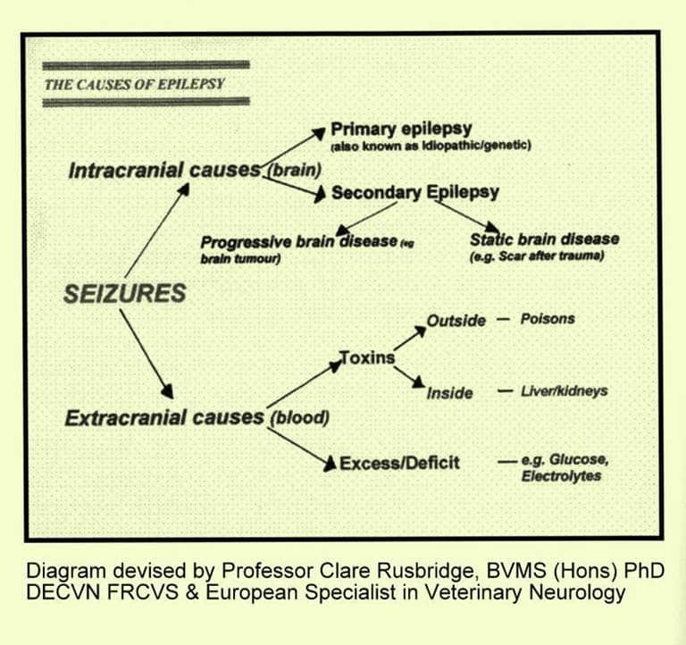
Epilepsy means repeated seizures due to abnormal electrical activity in the brain and is caused by an abnormality in the brain itself. However a fitting dog is not always an epileptic dog. Fitting or seizures can be caused by a variety of disorders (including poisons, metabolic disorders and brain tumours), with epilepsy being only one of them.
Epilepsy is recognised as an inherited condition (idiopathic epilepsy) in some breeds, and typically signs start between 6 months and 3 years of age.
Signs: Fits occurring during exercise are unlikely to be epilepsy. Epileptic fits usually occur when the dog is quiet and even when rising from sleep: the dog collapses, is unconscious and unresponsive, thrashes it’s legs, often froths at the mouth and can empty its bladder and bowels. It may also scream and moan loudly whilst fitting.
Action: Try to prevent self-injury to your pet, but do NOT attempt to pull its tongue out and never put your face near to a fitting dog; you may be bitten as your dog will not recognise you whilst he is fitting. Some restraint may be necessary, but letting your dog just lie on the floor is probably best, so do not try and move him unless he is in danger. Do not give stimulants. As he recovers he will recognise your voice, so talk to him all the time in a reassuring manner. Time how long the fit lasts and when he has recovered contact your vet; most fits last less than a minute – it just seems much longer. But if a fit does lasts for more than ten minutes or clusters of fits occur in rapid succession seek veterinary attention immediately.
On recovery, remove excess saliva and put the dog in a darkened room, keep quiet and warm. Keep a detailed record of your dog’s fits, and let the Breeder know once the diagnosis has been confirmed by your vet.
The Canine Epilepsy Support Group is a small charity set up in 1991 to offer practical and sympathetic support to the owners of epileptic pets, and the opportunity to talk to people who have learnt to live happily with an epileptic pet.
Their Advisory Panel includes Mr Francis Hunter, VetFFHom, MRCVS, Adviser on Homoeopathy, Mrs Sylvia Gulbenkian, BVetMed, MRCVS, Adviser on Acupuncture, and Professor Steve Dean, BVetMed, DVR, MRCVS, Adviser on Veterinary Legislation. The group also works closely with two Herbalists and a Holistic Therapist and they have set out to offer alternative options in addition to prescribed medication and veterinary care.Their aim is to help owners achieve normal, happy lives for their pets and are here to help and support you and your pet.: http://www.canineepilepsysupport.co.uk
The Phyllis Croft Foundation for Canine Epilepsy (PCFCE) was founded to bring comfort,support and information to the owners of epileptic dogs: http://www.pcfce.org.uk/
Update on Epilepsy Research at Canine Genetics Centre at Cambridge University.
Further update on the Canine Genetics Centre Epilepsy Study October 2024.
Many thanks to everyone who has supplied cheek swab samples from either affected or unaffected dogs. As well as the dogs from the UK, samples have been received from Utrecht University. The centre is aiming to collect DNA from around 200 dogs for each of the five breeds: ideally 100 cases and 100 controls so please keep samples coming in. They still have another approximately 9 months to collect samples and they’re hoping to reach this number and if they go beyond this it will be great as it’s a question of the more the better! So please keep sending samples and spread the word about the research.
The five IE cases and five controls for each of the five breeds are now being whole genome sequenced and they are finalising their analysis plans and computational scripts ready for the data to return in a few weeks – exciting!
The Canine Genetic Centre at Cambridge launched an epilepsy research project in March which included Irish Setters. Below is their latest call for samples from Irish setters which have been diagnosed with idiopathic epilepsy OR is over 8 years old AND unaffected by epilepsy.
Canine Genetics Centre at University of Cambridge
Earlier this year we told you about new funding that Dr. Sally Ricketts had secured, in collaboration with Linnaeus Veterinary and the University of Helsinki, to extend our epilepsy research into FIVE new at-risk breeds – the Beagle, English Springer Spaniel, Giant Schnauzer, Hungarian Vizsla and Irish Setter.
We have been actively recruiting samples from case and control dogs over the last 6 months ready to send for whole genome sequencing (WGS) and last month our spotlight was on the English Springer Spaniel where you were all amazing in your help, thank you! Now it’s the turn of the Irish Setter as we need more samples please
Do you have a setter that …
… has been diagnosed with idiopathic epilepsy
OR
… is over 8 years old AND unaffected by epilepsy?
If so, please contact the team to get involved and help with our research – https://www.canine-genetics.org.uk/research/epilepsy/

Epilepsy Research Study at The Canine Genetics Centre at the University of Cambridge
In September last year the Kennel Club Genetics Centre held a research day hosted by the University of Cambridge. For the three Breed Club Health representatives who attended the highlight of the day was the initial announcement of a new epilepsy study involving five breeds which includes the Irish Setter.
The breed clubs are actively supporting this new study and are eagerly waiting for information as it is released. For those of you who supported our initiative and supplied swabs to the University of Helsinki you’ll be delighted to know that they are one of the research groups involved.
Below is the latest update from the Canine Genetics Centre.
Epilepsy programme update
 Alongside our current genetic investigations of idiopathic epilepsy in the Border Collie and Italian Spinone, we are embarking on an exciting new study starting this year to investigate the genetics of epilepsy in five new breeds – the Beagle, English Springer Spaniel, Giant Schnauzer, Hungarian Vizsla and Irish Setter.
This two-year study is a collaboration with Linnaeus, the University of Helsinki and other veterinarians/research groups within our field, where we will utilise a newly developed approach, based on whole genome sequencing (WGS), to study the disease.
We are also keen to utilise the genomes of idiopathic epilepsy cases generated as part of the Give a Dog a Genome project, and so an arm of the study will extend to all of the breeds that participated in this project.
We are currently refreshing our website, so do look out for updates, and we hope to run a webinar later in the year to explain our study in more detail. More information coming soon as the study progresses!
Estimated Breeding Values (EBVs) are a tool that can help breeders make breeding choices based on indicators of the genetic risk of complex inherited disease, which is more accurate than by using an individual dog’s test score alone. EBV calculations use all BVA/KC hip and elbow screening data and pedigree information from the individual dog and its surrounding family, to more effectively determine the genetic risk that each dog will pass this disease to its progeny.
An EBV can only be calculated for a breed if enough individual dogs across the breed have been scored. EBVs will become available for more breeds, when there are enough scores within the breed to make the calculations.
An Estimated Breeding Value (EBV) evaluates the genetic value of an individual dog, in relation to the whole of the dog’s breed. These EBVs are intended to help breeders reduce the prevalence of hip and/or elbow dysplasia by more accurately evaluating genetic risk.
EBVs are usually updated quarterly to ensure that data on newly scored dogs are incorporated and that EBVs are provided for newly registered puppies. They are available on Mate Select.
Gluten-sensitive enteropathy in Irish setters
Ed Hall, MA VetMB PhD DipECVIM-CA, FRCVS
Emeritus Professor of Small Animal Medicine, University of Bristol
KC Breed Health Coordinator for the Irish setter
Summary
Gluten-sensitive enteropathy (GSE) is an intestinal disease that causes chronic diarrhoea and poor growth. It is caused by gluten found in wheat and by similar proteins in other related cereals (barley, rye, oats). First documented in a specific line of Irish setters, it has been bred out. However, it may still occur in setters sporadically and it probably occurs in other dog breeds although is rarely proven as proof requires repeated intestinal biopsy. It shares some similarities to coeliac disease in people, and an antibody test used for coeliac disease may be a future non-invasive test. Treatment simply requires feeding a gluten-free diet.
Nearly 40 years ago, I was the first person to demonstrate an enteropathy (i.e. a chronic intestinal disease) in Irish setters caused by a sensitivity to dietary gluten, a component of wheat. So-called Gluten-Sensitive Enteropathy (GSE) has, unfortunately, become synonymous with the breed even though the breeding from the line of affected setters ceased and dogs of other breeds can also be gluten-sensitive.
Irish setters affected by GSE developed intermittent or continuous diarrhoea after weaning and were typically underweight and stunted. Intestinal biopsies showed non-specific inflammatory changes which were resolved by feeding a gluten-free diet. My studies also showed that if gluten was excluded in the first six months after weaning, severity of the signs were reduced or delayed. Breeding studies showed a familial tendency and a possible link to specific tissue types, but the genetic basis was never elucidated. Using so-called permeability probes, small harmless molecules that can pass through a damaged intestinal lining, an underlying ‘leaky gut’ was demonstrated even in dogs reared on a gluten-free diet suggesting this was the underlying defect. Passage of gluten or its breakdown products could then stimulate the immune system via an abnormal route and induce inflammation with consequent gut dysfunction.
Parallels have been drawn between GSE in setters and a GSE in people known as coeliac disease. Curiously, coeliac disease is found most commonly in red-haired Irish people. There is a clear association with certain inherited tissue types as determined by molecules on the surface of cells (so-called HLAs) which are also important in donor-recipient matching for organ transplants. Coeliac disease can manifest as chronic diarrhoea at any age, but if it develops in childhood it affects growth as well. Historically, coeliac disease could only be diagnosed by intestinal biopsy repeated after remission on feeding a gluten-free diet and then again when challenged with a gluten extract. Similarly, proof of a GSE in setters required the observation of abnormal clinical signs and intestinal biopsy changes whilst the dog was fed gluten, clinical remission and resolution of the biopsy changes when placed on a gluten-free diet, and then relapse of signs and biopsy changes when challenged with gluten. Clearly this prolonged process with a need for three biopsies is not likely to be undertaken in general practice. However, many dogs with chronic diarrhoea do respond to exclusion diets and have a so-called Food-Responsive Enteropathy (FRE), but whether gluten is the inciting ingredient is never proven.
So what is gluten? Grains of wheat are composed mainly of starch but also contain two main proteins: glutenin and gluten. Glutenin is a polymer that makes dough sticky and allows bread to rise, whilst it is gluten that is the component that is harmful to coeliac patients. Other related cereals contain similar proteins: secalins in rye, hordeins in barley, and avenins in oats. Because of their similarity to gluten, people with coeliac disease are often also sensitive to barley and rye and sometimes to oats. Maize (known as corn in the USA) and rice are only distantly related to wheat and have proteins (zeins and oryzeins respectively) that don’t cross‑react in coeliac patients and are, therefore, safe to eat. Consequently, rice and maize are frequently used to provide carbohydrate in gluten-free diets for dogs. However, very confusingly, maize flour is sometimes labelled as “maize gluten”, but it does not contain true gluten. Soy, legumes and pulses are alternative carbohydrate sources that also do not contain proteins related to gluten. This, perhaps, has led to the fad of feeding “grain-free” diets but replacing cereals with legumes and pulses can predispose to the development of cardiomyopathy in some dogs.
As stated, GSE almost certainly occurs in many breeds, not just setters, but has never been proven because to have to perform three biopsies (affected–remission–challenge) is unrealistic. Indeed this process is no longer used to diagnose coeliac disease: an initial biopsy is paired with an antibody test to make the diagnosis. Antibodies to an enzyme called tissue transglutaminase (tTg) in the gut lining cross-react with gluten and are a marker for the disease. As yet nobody has tested for tTg antibodies in setters with suspected GSE. However, one research study found increased tTg antibodies in dogs with inflammatory bowel disease and alimentary lymphoma but did not then trial a gluten-free diet. Border terriers with paroxysmal movement disorders (abnormal, involuntary and often jerky movements, sometimes mistaken for epilepsy) have been shown to have increased tTg antibodies and do improve on a gluten-free diet.
Finally, successful treatment of GSE should simply be by feeding a gluten-free diet, i.e. avoiding wheat, barley, rye and possibly oats. Rice and maize should be safe to feed, and non‑cereal foods such as soy, legumes and pulses etc can be alternative sources of carbohydrate. Anecdotal stories of dogs affected with a GSE that also need steroids to control signs almost certainly didn’t have a GSE.
A gluten-free diet is often recommended for babies under six months of age as it was initially thought that early exposure to gluten predisposed to the development of coeliac disease. This is not current thinking, but baby food manufacturers still err on the side of caution and only produce gluten-free foods for babies less than six months of age. There is no evidence that feeding a gluten-free diet prevents GSE in setters, although it did appear to reduce the disease severity, or at least delay onset during the critical growth phase.
In conclusion, GSE has been reported in Irish setters but almost certainly occurs in other breeds. Proof is usually lacking, but undertaking a diet trial with an exclusion diet such as a single novel protein and carbohydrate diet or a hydrolysed diet, which are both gluten-free, is recommended.
- For Great British Bake-Off fans, rye contains very little glutenin which is why rye bread doesn’t rise much.
This
Final report on the Bloat survey conducted by AHT/KC
Received by Joint Breed Clubs’ Health committee
Animal data.
Surveys were completed for 1911 unique animals, from 1091 litters (mean 1.75 animals per litter), with 412 unique sires (mean 4.64 animals per sire) and 848 unique dams (mean 2.25 animals per dam). The breakdown of animals per litter is shown in table 1.
# in litter | count | % |
1 | 595 | 54.54% |
2 | 293 | 26.86% |
3 | 118 | 10.82% |
4 | 63 | 5.77% |
5 | 14 | 1.28% |
6 | 4 | 0.37% |
7 | 3 | 0.27% |
8 | 0 | 0.00% |
9 | 1 | 0.09% |
Table 1. Distribution of animals per litter in survey.
Of the 1911 unique animals for which surveys were completed, 1046 were female (54.74%) and 865 were male (45.26%). Table 2 shows cross-tabulation of neuter status with sex.
unknown | entire | neutered | |
13 (1.2%) | 733 (70.0%) | 300 (28.7%) | females |
7 (0.8%) | 667 (77.1%) | 191 (22.1%) | males |
Table 2. Cross-tabulation of neuter status with sex.
There was a large range of year of birth (yob), from 1991 to 2013 (the year of survey), with 93% of respondent animals born between 2000 and 2011.
Bloat data.
1657 of 1911 animals were reported as never having experienced an episode of bloat (86.71% of animals). 254 animals (13.29%) were reported as having experienced at least one episode of bloat at the time of survey, with details on 472 incidents of bloat supplied. Table 3 shows the distribution of animals and incidents by number of episodes reported.
Of the 472 reported incidents of bloat, just over half resulted in surgery or the death of the animal (50.85%), while 230 episodes were reported to have been resolved spontaneously, by management or medication (48.73%), see table 4.
episodes | animals | incidents |
1 | 165 | 165 |
2 | 39 | 78 |
3 | 8 | 24 |
4 | 5 | 20 |
5+ | 37 | 185 |
total | 254 | 472 |
Table 3. Distribution of number of reported episodes of bloat per animal.
Treatment / outcome | Count | Percent |
Don’t know | 2 | 0.42% |
Resolved itself | 83 | 17.58% |
Managed through diet/lifestyle changes… | 92 | 19.49% |
Managed with medication | 55 | 11.65% |
Surgery | 194 | 41.10% |
Died or put to sleep | 46 | 9.75% |
Table 4. Distribution of treatment/outcome of bloat epsidodes.
Data on multiple cases of bloat per animal were collapsed to generate a data field indicating whether the animal had ever experienced an episode of bloat (0=no, 1=yes) with an accompanying field specifying either age in days at time of the survey (where bloat field = 0), or age in days at earliest episode (where bloat field =1). This was repeated for bloat where treatment/outcome was specified as surgery or death/put to sleep.
Examination of the prevalence of animals experiencing bloat by yob revealed bias, with the prevalence tending to be higher in earlier yob (table 5), over the majority of the data (yob 2000-12). This bias is consistent with bloat having being more commonly observed in older animals.
The minimum age at survey for animals not having experienced bloat was 176 days, and the maximum was 5454 days (14.94 years). The mean (μ) and standard deviation (σ) age of animals at survey which had not experienced an episode of bloat was: μ= 2218.46 days, σ= 1161.05 days (6.08 years and 3.18 years respectively). The minimum age supplied for an animal experiencing its earliest episode of bloat was 61 days, and the maximum age for an animal experiencing its first episode was 4593 days (12.58 years). The mean and standard deviation of age at earliest episode were: μ= 1755.30 days, σ= 1116.38 days (4.81 years and 3.06 years respectively).
The minimum age at survey for animals not having experienced bloat resulting in surgery or death was 176 days, and the maximum was 5454 days (14.94 years), with μ= 2222.82 days, σ= 1164.11 days (6.09 years and 3.19 years respectively).The minimum age supplied for an animal experiencing its earliest episode of bloat resulting in surgery or death was 152 days, and the maximum earliest age reported was 4593 days (12.58 years), with μ= 1959.75 days, σ= 1097.18 days (5.37 years and 3.01 years respectively).
yob | Bloat with all treatments & outcomes | Bloat resulting in surgery or death | ||||
no bloat | bloat | prevalence | no bloat | bloat | prevalence | |
unknown | 2 | 1 | 33.33% | 2 | 1 | 33.33% |
1991 | 0 | 2 | 100.00% | 0 | 2 | 100.00% |
1992 | 2 | 1 | 33.33% | 3 | 0 | 0.00% |
1993 | 1 | 0 | 0.00% | 1 | 0 | 0.00% |
1994 | 0 | 1 | 100.00% | 0 | 1 | 100.00% |
1995 | 1 | 1 | 50.00% | 1 | 1 | 50.00% |
1996 | 1 | 3 | 75.00% | 3 | 1 | 25.00% |
1997 | 2 | 3 | 60.00% | 2 | 3 | 60.00% |
1998 | 1 | 3 | 75.00% | 2 | 2 | 50.00% |
1999 | 6 | 2 | 25.00% | 6 | 2 | 25.00% |
2000 | 62 | 14 | 18.42% | 62 | 14 | 18.42% |
2001 | 79 | 20 | 20.20% | 81 | 18 | 18.18% |
2002 | 90 | 26 | 22.41% | 93 | 23 | 19.83% |
2003 | 80 | 27 | 25.23% | 85 | 22 | 20.56% |
2004 | 114 | 25 | 17.99% | 120 | 19 | 13.67% |
2005 | 150 | 22 | 12.79% | 153 | 19 | 11.05% |
2006 | 143 | 27 | 15.88% | 145 | 25 | 14.71% |
2007 | 155 | 17 | 9.88% | 159 | 13 | 7.56% |
2008 | 170 | 12 | 6.59% | 173 | 9 | 4.95% |
2009 | 148 | 20 | 11.90% | 150 | 18 | 10.71% |
2010 | 179 | 15 | 7.73% | 186 | 8 | 4.12% |
2011 | 174 | 6 | 3.33% | 178 | 2 | 1.11% |
2012 | 92 | 6 | 6.12% | 95 | 3 | 3.06% |
2013 | 5 | 0 | 0.00% | 5 | 0 | 0.00% |
Table 5. Number and prevalence of animals experiencing bloat by yob
Genetic analysis.
A range of preliminary mixed models were run aiming to estimate the additive genetic variation in developing bloat. The proportion of phenotypic (measurable/observable) variation comprised of additive genetic variation is known as the heritability and describes the influence of genetics on the trait and how it will respond to selection.
Estimates of heritability from these preliminary models ranged from approximately 0.04 to 0.2, varying with the data and model used (data are routinely trimmed to minimise the number of classes of effect needing to be estimated; for example data may be limited to specific yob to avoid requiring the model to estimate effects of yob classes where data is scarce, such as 1991/2/3 etc, see table 5). Binary data (0/1), as used here, suffer a loss of precision in risk description in comparison to more continuous data (for example hip score ranges from 0 to 106), and this tends to lower the estimate of heritability (methods of adjustment are available). Thus, normally we might expect the true heritability to be higher than the estimates quoted above.
However, it was noted that the estimated effects of age from the analysis were extremely large, particularly when yob was also included in the model, and ran counter to previous reports (i.e. the models estimated that risk of bloat decreased as animals got older). This is a worrying observation, and is consistent with another bias in this data; that there appeared to be a higher prevalence of bloat in younger animals than in older animals. While we may surmise at the cause of this bias, there is no information on what actually causes it.
Therefore, while we have detected a signature of incidence compatible with genetics (estimated in the form of heritability), regrettably we do not think we can claim beyond doubt to have established that there is genetic variation in the likelihood of developing bloat. There are significant biases in the current dataset and the issue is whether these biases would either influence or produce the signature detected as an artefact. Although we feel this is unlikely, the presence of the biases described unfortunately prevent us from claiming to have determined risk of bloat is heritable in the Irish Setter.
Further work.
A further method of data analysis is currently being explored which may be particularly suited to data such as was collected from the survey. Survival analysis models the survival function from data of the age at which animals succumb to disease or death (and also the hazard function as the risk of disease at a particular age), and is a flexible way to incorporate censored data (i.e. animals unaffected or still alive at time of survey). Survival Kit software has been tailored towards animal breeders and allows the estimation of random effects (i.e. genetic variation). Work on using Survival Kit software and running survival analysis is being undertaken.
While we are unable to state categorically that bloat is heritable in the Irish Setter, the evidence does appear to indicate that genetics could play an important role. Given the prevalence of bloat in this breed, and the concerns of breeders and owners, it may be useful to attempt to ‘extend’ the survey, by building and maintaining a database of details of dogs affected and unaffected by bloat. As the amount of data increases, it becomes easier to reduce the effects of bias seen in the current data set, for example if enough data were available analysis could focus solely on older dogs, reducing the influence of age (and providing a more reliable indicator of ‘lifetime’ risk). Assistance in this undertaking may be available from the Kennel Club.
Breeders may also wish to explore the idea of taking and storing DNA samples from both affected and unaffected dogs alongside a database of information as suggested above. While it is highly unlikely that a mutant variant of a single gene is solely responsible for the development of bloat, genetics could certainly have an important role, as stated above. As such, taking and storing DNA samples may bequeath the breed in future with an extremely useful resource to begin to track down regions of the DNA and maybe even genes which are responsible for increasing or decreasing the risk of developing bloat. Because there are almost certainly multiple risk factors for bloat, some of which might be genetic, it will be necessary to collect DNA from large numbers (many hundreds) of dogs that have suffered at least one attack of bloat as well as DNA from a similar number of old dogs that have never had bloat before a genetic study is likely to be successful.
January 2017
…………………………………….
INTERIM REPORT
KC/AHT Bloat Survey.
The AHT/KC survey of Irish setters last year, with financial support of the JISBC, was aimed primarily at gathering evidence to prove that bloat is heritable. If proven, DNA samples could then be evaluated for possible genetic markers. However, it was always understood that it was very unlikely to be a simple inheritance (i.e. single gene mutation) pattern. Whilst there may be genetic susceptibility, environmental factors are likely to play a significant role.
Regrettably, although the results raised a suspicion that there is an inherited component to the condition, confounding factors prevented the results reaching statistical significance and therefore proof. The number of respondents and the fact that some of the dogs alive at the time of the survey may go on to develop bloat later in life confounded any clear conclusion.
The recommendation must therefore be that the breed prospectively collects DNA samples from dogs that suffer bloat. Whilst this outcome is disappointing, it is recommended that we work with the AHT to collect DNA samples and pedigrees of dogs when they suffer bloat confirmed by surgery or post mortem.
As you will be aware, the survey also gathered information on a number of other conditions. This data has not yet been analysed, but I have asked Tom Lewis (KC, formerly AHT) to analyse results pertaining to megaoesophagus and epilepsy, where it is more likely that heritability can be proven.
Ed Hall
Chairman, Joint Irish Setter Breed Clubs Health Committee
22/3/2015
In 2004 the Kennel Club and British Small Animal Veterinary Association joined with the Animal Health Trust to carry out a survey of pedigree dogs in UK. A questionnaire was devised and sent to owners to try and identify which health conditions were present in each breed. The questionnaires were circulated by breed clubs to their members.
The questionnaire was divided into different sections with questions on the health of the owner’s dogs, breeding, causes of death and birth defects in any puppies.
Where the breed response was 15% or greater, breed clubs received detailed feedback on the results which the Kennel Club felt should help with the recognition and control of important conditions in specific breeds. It further felt that Data gathered would be the baseline against which the success of future control schemes can be measured.
The Irish Setter breed response was 24%.
The following link is to the results of this survey:
http://www.thekennelclub.org.uk/media/16534/irish%20setter.pdf
Our breed health and conservations plans (BHCPs) use evidence and data to help us understand the health issues found in each pedigree dog breed. These plans help breeders and owners identify health and welfare problems and use information, health tests and health schemes to avoid passing on those problems to future puppies. They also support and provide breeders with tools and specialist expertise to help manage genetic diversity, understand the impacts of close breeding, and find the best ways to preserve the population of their breed.
Working together for the breed
We’ve worked with breed clubs and breed representatives to gather all available evidence to help us determine the priority concerns for the breed and decide how we can work together to manage and reduce these problems.
The full evidence base is available at the discretion of the breed clubs, however if you would like to seek access to the full report, please contact our health team.
More about health
If you have any concerns about a particular health condition in your breed then you may wish to speak to your vet or you could contact your breed health co-ordinator.
Breed health co-ordinators are individuals working on behalf of breed clubs and councils who are advocates for the health and welfare of their chosen breed. They act as a spokesperson on matters of health and will collaborate with The Kennel Club on any health concerns the breed may have.
To contact your breed health co-ordinator please email:
Breed watch
Category 1
Currently no points of concern specific to this breed have been identified for special attention by judges, other than those covered routinely by The Kennel Club’s breed standard.
Breeding restrictions
There are a number of The Kennel Club’s rules and regulations that may prevent a litter from being registered, find out about our general and breed specific breeding restrictions below.
More about breeding
With effect from 6 January 2025, imported Irish Setters and litters from overseas sires will only be eligible for Kennel Club registration if they are proven to be clear or carriers of canine leukocyte deficiency (CLAD) and progressive retinal atrophy (PRA rcd1), or if they have hereditary clear status.
Identified carriers may be used for breeding, provided they are only mated to an Irish Setter that is either hereditarily clear or DNA-tested clear of CLAD and PRA (rcd1).
For litters with overseas sires, the sire must be proven to be either clear or a carrier of the conditions.
This scheme has been introduced at the request of the Breed Club(s) to safeguard the UK Irish Setter population from the introduction of these conditions.
The breed health and conservation plans ensure that all health concerns are identified through evidence-based criteria which is compiled into a single document. The Kennel Club has gathered all available health information and data about each specific breed which is used as a basis to collaboratively create a strategy to tackle the health priorities of that particular breed.
Click below to load pdf file
This information is now held on KC website and can be accessed through the health test results of individual dogs. https://www.thekennelclub.org.uk/search/health-test-results-finder/
The condition consists of a degeneration of the nerves, which stimulate the muscles of the voice box (larynx). Paralysis of the larynx is quite common in elderly dogs, especially males, and although the Labrador, Irish Setter and Afghan Hound seem to be particularly susceptible, practically any breed in the middle weight range could be involved.
Signs may go unnoticed because owners expect elderly dogs to slow up and huff and puff a bit when exercising. One or more of the following are the most frequent signs of laryngeal paralysis:
- Noisy laboured breathing
- A moist retching cough
- Changed bark
- Reduced exercise ability
- Episodes of extreme breathing difficulty, especially when exercising in hot weather.
Collapse and death can occur if the loose vocal folds block the airway completely.
If you believe your pet has this problem it is necessary to see your vet to get the diagnosis confirmed. Treatment is by operation to fix the voice-box in a safe position. In spite of the age of many dogs subjected to surgery, the results are generally excellent.
Following the operation, the dog may be hospitalised for between two to four days, although dogs that bark excessively may be sent home earlier if there is concern they will tear the stitches.
Diet and exercise should be modified for the first six weeks after surgery as advised by your vet.
Although an immediate improvement of the respiratory distress may be evident, the full benefits of the surgery will not be seen for a couple of weeks, when the internal swelling has gone.
Most dogs cough to clear their throats to begin with, following ‘tie back’ surgery. This may be quite frequent in the first week or so, particularly after eating or drinking. The coughing should get less frequent, although a few dogs can cough once or twice a day indefinitely.
Lungworm or French Heart Worm
Lungworm (Angiostrongylus vasorum) has only become a potential problem in UK in the last 15 or so years and affected dogs are now seen as far north as Scotland. It is generally felt that our warmer climate is the reason for its spread. It has a complicated life cycle and dogs eating the snail or slug is part of the cycle. In dogs, the worm usually lives in the blood vessels passing from the heart to the lungs, but it can migrate to other sites including the eyes and brain. It does not affect humans and cannot be transmitted to you through your dog.
Dogs that are at risk of this parasitic worm are those that either eat slugs or snails deliberately,or eat grass and accidentally ingest small slugs and snails. Younger dogs seem to be more likely to get infected but it is not unknown for older dogs to suffer.
Vets will advise lungworm treatment as part of your dog’s health regime because if it is not treated it can lead to death. If you have one dog that is affected it is sensible to treat all your dogs.
Symptoms include:
Coughing
Breathing problems
Weight loss
Vomiting
Diarrhoea
Persistent bleeding from cuts
Depression
Not wanting to exercise
Weakness
Paralysis
If you know you have snails or slugs in your garden:
Don’t leave water bowls outside
Don’t leave toys and chews outside
Be particular about removing dog faeces daily
Don’t assume your usual worming tablets treat lungworm
Ask your vet about the treatment that is available and make sure your dog is
treated regularly.
The link below takes you to the Kennel Club article on lungworm
Go to the site below to see a cartoon video of the life cycle of the lungworm
http://www.youtube.com/watch?v=gHgmIc4Vbrw&feature=player_embedded
The oesophagus is the muscular tube that takes food from the mouth to the stomach. This is done by waves of muscular contractions, called peristalsis, which push the food along the tube.
Megaoesophagus (MO) refers to a large, flabby oesophagus which makes it difficult or impossible for food to reach the stomach because the peristaltic action does not happen as it should, probably because the nerves are not functioning properly. Food cannot enter the stomach normally, but instead simply sits in the enlarged oesophagus and is eventually regurgitated.
Some cases of MO in Irish Setters are congenital, i.e. present at birth, but it may not be noticed that the pup has any problem until it is weaned when he will regurgitate food through the mouth and maybe fluids through the nose. It may cough and make gurgling, rattling sounds. An affected pup generally will not thrive and will probably be smaller than his littermates.
MO can also be acquired later in life (about 4 years of age onwards) with similar clinical signs and poor prognosis.
The signs of MO are as follows:
Regurgitation may be considered the most typical sign of MO. Weight loss with possible muscle wasting and a failure to thrive with a general weakness are common. Increased swallowing motions with excessive drooling and dehydration are possible. A ravenous appetite but with stunted growth or weight loss are usual, as is coughing, difficulty in breathing and pneumonia.
Regurgitation is different from vomiting: Vomiting occurs when the contents of the stomach are expelled by muscular contractions of the abdomen. Regurgitation is purely the return of food that has not reached the stomach and, as such, retching does not happen. As it happens very quickly and with little effort littermates or mum may clean up the results before the breeder realizes it has happened.
Dogs with MO may not exhibit all of these signs, or even any of them to a significant degree. Sometimes the only signs may be repeated bouts of aspiration pneumonia, or a wet cough that fails to clear up. Some pups with congenital MO can grow out of the disorder and go on to enjoy a normal quality of life but others will be significantly affected and need careful food management for the rest of their lives. If the problem is severe, however, the pup will not be able to get enough food and will have to be euthanased. Acquired disease in adult dogs never resolves.
A definitive diagnosis can be obtained by giving a barium meal. In a normal pup, the barium will move into and through the stomach, but in the dog with MO, most of it will be seen collected in the oesophageal pouch in front of the stomach.
Another congenital reason for regurgitation is a vascular ring anomaly such as persistent right aortic arch. Foetal blood vessels that should have disappeared at birth create a fibrous band that constricts the oesophagus. This causes the oesophagus above the constriction to expand as the food cannot pass through the constricted area. If caught in time, the vascular ring can be cut and the oesophagus often returns to normal. Delaying surgery may cause irreparable oesophageal damage.
Oesophageal dilation and vascular ring anomalies are both believed to have a hereditary component because there is a breed disposition and a probable family predisposition.
If you believe your pet has MO then you will need veterinary advice. If confirmed it is important to let your breeder know as well as the secretary of one of the breed clubs as information is being collected on the problem.
Follow the next link to an excellent article on MO with clear X rays of a dog without MO and one with MO. There is also a very clear visual of a dog with MO trying to eat
http://www.marvistavet.com/html/body megaesphagus.html/
Sometimes MO doesn’t happen until later in life, maybe through trauma or being associated with other health problems but this form is not generally a problem with Irish Setters.
Can you help?
If you breed a litter with MO or your puppy is diagnosed with MO please let the Breed Health Committee know so we can monitor the prevalence in the breed.
As a breed we are used to submitting DNA samples from our dogs either for specific disease testing such as PRA-rcd1, PRA-rcd4 and CLAD, or for sending DNA for research purposes such as epilepsy.
There are, however, other uses for DNA, such as DNA profiling and parentage testing. The article below is written by Dr June Swinburne – Director of Animal DNA Diagnostics Ltd – in which she covers some of the advantages in adopting routine DNA profiling for our dogs:
If you value accurate pedigrees for your dogs and the wider breed population, then the scientific validation of their accuracy should be considered; undoubtedly errors exist in current pedigrees, whether accidental or not! When planning breeding the accuracy of pedigrees is valuable, but if at any point an inherited disease requires investigation this becomes crucial in investigating the basis for the inheritance, and which individuals are at risk. I hope that the following article will provide a basis for understanding how DNA profiling works, and give food for thought and discussion!
Uses of a DNA profile
There are several uses for DNA profiling. These include:
- To enable parentage verification and pedigree validation
- As a research resource if new diseases emerge
- To obtain a unique identification of the animal at the time of disease testing
- To obtain a unique identification of the animal in the event that he is stolen or lost
A DNA profile consists of data from a selection of genetic ‘markers’. There are thousands of genetic markers in a dog’s DNA, and these are of several different types; the ones we use for profiling are known as ‘microsatellites’. Typically a DNA profile will consist of data from 13 – 19 microsatellites. The microsatellites chosen for use are called a ‘microsatellite panel’.
It is extremely likely that the DNA profile from any one dog is completely unique. For this reason it is similar to a fingerprint, and indeed a DNA profile is often referred to as a ‘DNA fingerprint’. The only exception to this uniqueness would be between identical twins, which are genetically identical.
Different laboratories around the world could choose different selections of microsatellites for the panel that they use, however usually there is a core of markers common to most labs. These core markers are decided by scientists at the conferences held by the International Society of Animal Genetics (ISAG), which take place every second year. Here scientists working in the field discuss the pros and cons of the different microsatellites that they have used and decide which ones work best. The panel of markers which Animal DNA Diagnostics Ltd uses is a commercially available panel consisting of 19 markers.
Parentage verification
The basis for the use of microsatellites in parentage verification is that they vary in length. Each animal has two copies of each microsatellite, one inherited from each parent. In its most basic form, parentage verification checks that the copies inherited by the puppy are also present in the presumed parents – if they are not then the puppy cannot be from these parents. In order to perform parentage verification DNA profiles from the dam, all possible sires and the offspring are required.
This is much easier to understand in an illustration:

The illustration above is for one microsatellite. As shown the dam shares copy H with the puppy (‘Progeny’), and Sire 1 shares copy D with the puppy, so for this microsatellite the inheritance agrees with Sire 1 as the father. In contrast Sire 2 is excluded as the father as he shares no copy with the puppy. Just by chance a potential father could share copies with the puppy, and it is for this reason that more than one microsatellite is examined. As the number of microsatellites examined is increased, the chances of a male sharing copies of all microsatellites with a puppy which is not his offspring decreases dramatically. The panels in use for parentage testing today (consisting of 13-19 microsatellites) can reduce this chance to negligible. The probability that the qualifying male is the father is therefore very high.
There is one situation where care must be taken. This is in the case where either of two closely related males could be the father. In the vast majority of cases parentage verification will distinguish which of these males is the sire. In rare cases paternity is not conclusively determined with the primary marker panel and further markers are required. It is important that all males which could be the sire are tested; this is especially important where potential sires are closely related.
We have focussed on questioning which male is the sire of the puppy, but the analysis does also confirm that the presumptive dam is in fact the mother; in rare cases a puppy is paired with the incorrect mother and this will be detected.
Sample collection
A DNA profile is obtained from a sample of an animal’s DNA. This DNA sample can be extracted from a number of different sources including, among others, a blood sample, a mouth swab or hair roots. The DNA obtained from each of these sources is, to all intents and purposes, the same and will not change during the animal’s lifetime. So a DNA profile obtained from a puppy would be identical to that obtained from the mature dog.
For dogs, the most usual source of DNA is from a buccal swab – that is a sample of cheek cells taken from swabbing the inside of the animal’s mouth. The aim of a buccal swab is not, as is often thought, to obtain a sample of saliva, but rather to collect loose cheek cells which are constantly being shed inside the mouth; these are easily collected with a cotton swab or collection brush by rubbing this firmly against the inside of the cheek.
One drawback to buccal swabs is that they can potentially be contaminated with DNA from other dogs. This is more likely in un-weaned puppies who may have their mother’s DNA inside their mouths. Young dogs may also have DNA from their companions inside their mouths. For this reason it is preferable for dogs to be separated from their companions for at least one hour before sampling. In addition, to prevent contamination of the sample, dogs should not have access to food for 2 hours before sampling.
Once taken the swab should be left to dry thoroughly before being sent to the laboratory. Swabs do not need to be refrigerated, and can be sent within the UK in the normal mail in suitable packaging.
Once the swab arrives the DNA will be extracted and stored frozen. DNA can be stored frozen indefinitely, but the quality of the sample cannot be guaranteed for extended periods.
Information submitted with the sample
Generally the KC registered name, KC registration number, date of birth, sex, microchip number, sire and dam’s names and registration numbers are submitted with the sample. Sampling may occur before KC registration, but ideally not before microchipping, as it is essential that the dog can be identified.
Avoiding sampling failure and error
The most usual problem encountered with buccal swabs is that the swabbing is not performed adequately and that there is little or no extracted DNA. This can only be avoided by the owner obtaining a suitable sample by preparation and careful swabbing.
In addition, the possibility of mix-up does arise where several dogs are being swabbed in the same session, and swabs become mixed up. This is particularly a problem when litters of puppies are sampled which are of similar appearance. The owner must take great care that the sample is labelled correctly and can be matched with the correct puppy.
A more exacting sampling procedure can be put in place by requiring that the sample is taken by a vet. This could be combined with checking other identification such as a microchip or tattoo, the details of which can be attached to the DNA sample. This approach is used in the registration of Thoroughbred racehorses, where foals are micro-chipped at the same time that a DNA sample is taken; parentage verification is mandatory for their entry to the studbook. The use of a veterinary surgeon does however add to the costs. An alternative could be that the samples are taken by or confirmed by a third party, such as Breed Society officials, often at a breed show or a clinic specifically held for DNA sampling.
The possibility must be considered that owners may purposefully submit samples from the wrong puppy. This could be profitable for an owner where parentage of a puppy could add to his value; a sample from a legitimate puppy from the preferred parents could be submitted in place of the faked puppy’s sample. One could also imagine scenarios where false submission could be used to avoid KC restrictions on litter numbers. Although these dishonest practices could be imagined, in practice these should be rare as parentage testing in the next generation would uncover any anomalies.
Great care is taken in the laboratory to avoid sample mix-up. Procedures are put in place to ensure that this is avoided.
Data storage
A database will store the DNA profiles, and perform parentage verification. In it’s most basic form the parentage verification analysis would simply ask the question “Does the male(s) suggested as the sire of this puppy qualify as the father?” As the database builds up increasing numbers of profiles, the additional question could be asked “Do any other males on file qualify as the sire?” and consequently a more accurate probability can be assigned to the parentage assignment.
This article was written by Dr June Swinburne when she was Director of Animal DNA Diagnostics Ltd
Porto-systemic shunts (Liver shunts)
Professor E J Hall
A porto-systemic shunt (PSS) is an abnormal vessel that bypasses the liver so that blood which would normally drain from the intestines (via the portal vein) to the liver is ‘shunted’ directly into the general circulation. This causes significant ill health because of toxins from the gut reaching the brain. Ideally the shunt is surgically corrected.
Shunts are being recognised with increasing frequency in pedigree dogs (and occasionally in pedigree cats). They are most common in giant and toy breed dogs. Occasional cases have been seen in Irish setters although fortunately this is not (yet) a well known problem in the setter world.
We know that shunts are an inherited condition in the Irish wolfhound, but because of the prevalence in other specific breeds we suspect it is inherited in most cases. The exact genetic defect is not known yet, but work is underway in the USA. However, the mode of inheritance is not simple and parents and littermates may not be affected. However, breeding from parents that have produced affected offspring, or from affected animals cannot be recommended.
This article was written at the request of the Breed Club Health Coordinators with the aim of both raising awareness of this condition, so that cases are recognised and successfully treated, and ensuring appropriate measures to control breeding are applied.
This article draws on a client FAQ sheet given by the author to owners of affected dogs referred to Bristol Veterinary School.
What causes a shunt ?
This is a congenital problem, but although a patient is born with the PSS, signs usually only begin to develop weeks or even months after weaning, as the protein content of the diet increases. It is likely an inherited condition and breeding from affected animals is not recommended.
What does a PSS do to the animal?
A PSS can have a number of consequences:
1.Toxins [including ammonia (NH3)] produced by bacterial fermentation of protein in the intestines are not filtered by the liver and affect the brain. Variable neurological signs of ‘hepatic encephalopathy’ may occur e.g. restlessness, intermittent blindness, aimless wandering, head pressing, disorientation, increased thirst and even fits (seizures) and coma in severe cases.
2. Nutrients are not metabolised by the liver, which remains small. This can lead to stunting of the animal.
3. If the liver fails to produce adequate blood proteins, fluid may accumulate in the abdomen (‘ascites’) giving a pot-bellied appearance.
4. Sometimes the abnormal liver function leads to formation of stones in the kidneys and/or bladder and signs of blood in the urine or even obstruction.
5. Occasionally bacteria escape from the gut and, having evaded the liver, enter the circulation causing periods of ill health and raised temperature.
Where is the shunt ?
There are many anatomical variations on a theme, but in general there are two main types:
1.Intra-hepatic – the vein draining the intestine passes through the liver without dividing. This arises most frequently from failure of a vessel normally only present in the foetus to close. It is most commonly seen in giant breed dogs, and is a surgical challenge to correct.
2.Extra-hepatic – the shunt completely bypasses the liver and enters the general circulation directly via one of several possible routes; porto-caval is the most common type. Extra-hepatic shunts are more amenable to surgical correction.


What is the ideal treatment ?
In an ideal world the PSS is tied off (ligated) surgically, and this can be curative. The success rate varies between 50 and 85% depending on the type of shunt and surgical expertise. At Bristol Vet School, we can also now attempt to treat intra-hepatic shunts by placing an occluding coil via a venous catheter. There is still a risk with this new procedure but even riskier open surgery is not required
In some cases, ligation is not possible, for either medical or financial reasons. These patients are managed medically to control the signs of hepatic encephalopathy. Medical treatment merely reduces the production of toxins and does not correct the shunt.
What can go wrong ?
The aim of surgery is to completely close the shunt. Regrettably it is not always that straightforward:
- The shunt may be impossible to find
- There may be inadequate veins going to the liver (or even none) so that complete closure of the PSS causes excessive back-pressure on the intestines. In mild cases this may cause temporary accumulation of fluid (ascites). In severe cases it can lead to death of the patient, and so the surgery has to be reversed.
- There is a risk of serious haemorrhage, especially with intra-hepatic shunts, which may have to be dissected free of surrounding liver tissue. Placement of a coil by venous access is less risky but not widely available.
If the shunt is found but complete closure is not possible, a partial ligation may be performed. Alternatively a sterile cellophane band placed around the shunt, in order to cause scarring and gradual closure to allow time for the vessels to the liver to regrow.
What is medical management ?
The aim is to reduce intestinal production and absorption of toxins such as ammonia, and so reduce signs of hepatic encephalopathy. Medical treatment is indicated for:
- For short-term stabilisation of patients before surgery
- Patients where ligation of the shunt fails because of a lack of normal vessels going to the liver to cope with the revised blood flow
- Patients where surgery is declined for whatever reason.
There are three lines of treatment
1.Dietary management
A restricted protein diet with carbohydrates as the main energy source should be fed. Veterinary diets such as RCW Hepatic Support or Hill’s l/d are suitable. Alternatively a home-prepared diet consisting of equal parts of boiled rice, pasta or potatoes with low-fat cottage cheese may be fed. IIf blood proteins are low, protein should not be restricted severely, and other methods must be used.
2. Lactulose
This synthetic sugar is a laxative that helps remove the intestinal contents rapidly before significant fermentation occurs. It also decreases the absorption of ammonia. The effect is quite variable, and the dose has to be tailored until the patient produces 2-3 soft motions per day.
3.Oral antibiotics
These help reduce the number of ammonia producing bacteria in the gut lumen.
Treatment is tailored by trial and error to each individual patient until signs of hepatic encephalopathy are controlled. Mild cases may do well on dietary management alone, whilst severe cases may require all three medications.
NB. Cases of PSS must be referred by their vet to other centres offering surgery (including Bristol Veterinary School); owners cannot make arrangements directly.
If your Irish Setter is diagnosed with a liver shunt, which is very rare in the breed, then please let your breeder know.
One of the health problems people associate with Irish Setters is PRA (Progressive Retinal Atrophy) which is a term for several different forms of hereditary conditions which lead to blindness and which is found in many breeds of dogs. This was a major problem for the breed in the 1940’s and 1950’s and was the greatest threat to the breed. This eye condition leads to gradually worsening vision and eventual total blindness in both eyes. The condition is hereditary and is carried by a simple autosomal recessive gene.
The breed now has a DNA test for PRA rcd 1 mutation. Since the KC started its open register in 1995 no dogs that have been tested since then have been diagnosed with PRA rcd1 in UK. This is an early onset form of the disease, puppies typically being diagnosed from about 6 weeks and being totally blind by about 12 months, with night blindness being noticed first. Owners may notice that the dog is bumping into things in the dark or be unwilling to go outside.
By using the DNA test effectively this particular PRA is no longer a problem for Irish Setters in UK and since January 1st 2010 the Kennel Club will only register Irish Setters that are proven to be clear of PRA-rcd 1 (Progressive Retinal Atrophy), or hereditarily clear of PRA -rcd1 e.g. both parents are clear. This includes imported Irish Setters as well. Do not buy a puppy unless both parents are clear. This is clearly shown on the KC registration papers of the puppy. Remember both parents need to be clear not just one of them.
Why bother to have a PRA rcd 1 clear puppy?
There are several reasons why this is important; the first being that if a pup is clear then it will never get PRA rcd1 and if you decide to breed then its puppies will never get the problem. Responsible breeders have worked very hard to eradicate PRA rcd1 from the breed in UK and want to keep it that way. Also, although you may not be thinking of breeding from your pet at the moment, you may change your mind later and unless both parents are clear from PRA rcd1 then the puppies cannot be registered with the Kennel Club.
In 2014 we were advised that an Irish Setter in mainland Europe had been confirmed as being PRA-rcd1 affected and was going blind. In UK it is very easy to become complacent and believe this problem no longer exists but it obviously does.
Late onset PRA Blindness
Although PRA rcd1 is no longer a problem it is suggested you have your dog’s eyes tested at an eye clinic every two years, as there are other forms of PRA being identified.
Late Onset PRA (LOPRA) which, as the name suggests, does not show until the dog is older, has been identified in the breed. A mutation known as rcd4 has now been found, and a DNA test is available (NB. rcd2 and rcd3 are mutations found in other breeds). Time of onset of blindness is variable but typically later in life.
Recently a mid-onset PRA has been identified, with clinical signs of PRA developing in middle age. The genetic mutation has not yet been identified and research is ongoing.
Please look at “Spotlight on” to see the latest announcements about PRA rcd4 which is the mutation recently identified and for which a DNA test became available from 1st August 2011.
If you believe your Irish Setter has vision problems consult a veterinary ophthalmologist for a diagnosis of PRA. If it is confirmed then let the health representative of a breed club and your breeder know immediately. They will be able to advise you what to do next as a DNA test will be needed to confirm if it is PRA rcd4.
Any Irish Setter with suspected sight problems can have DNA testing free-of-charge if the sample sent to the AHT is accompanied by a certificate from a veterinary ophthalmologist confirming PRA (Progressive Retinal Atrophy) which is neither rcd1 or rcd4.
Pyometra is an infection of the uterus which needs immediate veterinary attention. If not treated it will be fatal. Any unspayed bitch can be affected although it is usually found in older bitches and signs are generally noticed 6-8 weeks after her season. If a bitch has had puppies it will stop her from getting a true pyometra but she can still suffer a uterine infection.
Causes.
When the bitch is in season there are hormonal changes to the uterus which is preparing itself for puppies. She has a bloody discharge, which is normal, and her cervix, which is normally closed, opens slightly to release this discharge before mating takes place. The open cervix can allow bacteria to enter the uterus which has become an ideal breeding place because repeated hormonal changes during each season have altered its lining.
Open or closed pyometra.
Sometimes the uterine cervix remains slightly open and this is called an open pyometra and is usually easier for the owner to notice as there will be a foul smelling discharge which is totally unlike her usual discharge. If the uterus closes completely this is a closed pyometra and there is no obvious discharge. This makes it more difficult for an owner to realise there is a problem and by then the infection could be severe. This is why it is really important to watch your bitch closely 6 – 8 weeks after her season to see if there are any symptoms.
Signs.
In both open and closed pyometra some of the following may be noticed but don’t wait to see them all. Sometimes all you notice is that your girl is “off colour” and you can’t be certain as to what is wrong. That is enough to make an appointment with your vet, especially if it is 6- 8 weeks after her season.
- Listless and depressed
- Drinking a lot of water
- Loss of appetite
- Distension of the stomach: particularly in a closed pyometra
- Vomiting
- Diarrhoea
- Fever
- Dehydration
With an open pyometra you may also see:
- Cleaning herself constantly under her tail
- Pus on her bedding and on her tail
- Foul smell from the pus
Treatment.
The usual and most effective way to treat pyometra is surgery to remove the womb and ovaries and this is probably what your vet will recommend. The spread of infection during the operation is a great worry and it is likely that antibiotics will also be given. However, there is a new treatment that is now available which means an operation may not be necessary.
It is important that you watch your bitch closely 6 – 8 weeks after her season as that is the time when you will see the signs. The earlier she is treated the better the chances of her survival.
This link is to Wikipedia which has photographs but it is not for the squeamish:
http://en.wikipedia.org/wiki/Pyometra
The following is a link to a scientific paper Canine Pyometra: Pathogenesis, Therapy and Clinical Cases presented by Prof. Stefano Romagnoli, University of Padua, Italy
http://www.vin.com/proceedings/Proceedings.plx?CID=WSAVA2002&PID=2686
Royal
https://www.rvc.ac.uk/research/projects/epilepsy-in-companion-animals
Autoimmune diseases are encountered frequently in dogs although thankfully the problem is not seen frequently in Irish Setters.
RVC researchers have launched an innovative new research project called the RVC ImmunoRegistry. This detailed investigation about autoimmune diseases in dogs involves collecting data in real time over one year after diagnosis to find out how affected dogs respond to treatment.
All participating dogs must:
Reside in the UK (are there any restrictions on age of dog or neutered status etc)
Have received a diagnosis of one of the conditions below within the past 10 days
– IMHA – immune-mediated haemolytic anaemia
– ITP – immune thrombocytopenia
– IMPA – immune-mediated polyarthritis
– SRMA and steroid-responsive meningitis arteritis
If you believe you have a recently diagnosed dog that would be eligible for this study, please visit the link below:
http://www.rvc.ac.uk/research/projects/rvc-immunoregistry-research-into-canine-autoimmune-diseases
The loss of a beloved companion can be the worst part of owning a pet. And when that tragedy is because of an unexpected and premature death through illness, the feelings of loss and grief are magnified. At such times one often feels that one’s pet has suffered enough and burial or cremation will allow closure. At that time the thought of a post mortem examination is anathema to many owners.
Regrettably, nothing is ever going to bring one’s pet back, but some solace can be gained by finding out the exact cause of an unexpected death. However, even more importantly, other dogs can benefit in the future. Whilst a post mortem examination clearly cannot help your pet, it can help vets understand diseases and ultimately learn something that may make a difference to the next dog to suffer the same condition. In addition, dogs specifically donated to Vet Schools provide essential training for tomorrow’s vets.
Yet even knowing the benefits that a post mortem examination can bring, many still will not contemplate this for fears that their pets will be experimented upon and that their organs will be retained for research. As someone who works in a Vet School, I can assure you that this does not happen; animals are treated with the same respect as if they were alive.
Post mortem examinations undertaken for legal cases do incur significant costs, and your local practice may charge for the examination. However, many Vet Schools will provide a post mortem service at a reduced cost or even for free for donated animals; they may only charge if you want a full written report. Unfortunately after a post mortem examination, release of the body for burial at home is not allowed because of the risk of infections; but individual cremation with the return of ashes can be arranged, although this service does incur a cost.
Finally, the JISBC Health Committee has become aware of a number of cases of unexpected sudden death in Irish setters due to internal bleeding. Post mortem examinations will be essential to identify the prevalence of this emerging problem, and to investigate its cause, with the ultimate aim of finding a solution. Recently reported cases have occurred in Scotland, and Dr Tennant at the Scottish Agricultural College has said they are willing to provide post mortem examinations at minimal cost on setters that have died suddenly.
So whilst saying goodbye is always hard, please think whether some good can come out of the loss of your pet by considering a post mortem examination.
Prof Ed Hall, JISBC Health Coordinator
Anecdotally it is known in the breed there is a chance that a bitch that has been spayed will develop spay incontinence and this is supported by the following research:
Incontinence is mentioned in the KC Breed Health Survey 2004 and appears in the list of the most common specific conditions reported.
https://www.thekennelclub.org.uk/media/16534/irish%20setter.pdf
The key findings included in more recent research published in September 2017 indicate:
- Urinary incontinence affects 3.14% of bitches overall, but affects over 15% of bitches in some high-risk breeds
- High-risk breeds include the Irish Setter, Dobermann, Bearded Collie, Rough Collie and Dalmatian
- Bitches weighing above average for their breed had 1.31 times the odds of urinary incontinence compared with bitches weighing below average
- Older bitches were also predisposed, with bitches aged between 9 and 12 years old having 3.86 times the odds compared to younger bitches
- Neutered bitches had 2.23 times the odds compared with entire bitches
RVC veterinary epidemiologist and VetCompass researcher Dr Dan O’Neill said: “This urinary incontinence study has uncovered dramatic breed predispositions that have previously been hidden to vets and owners. Overall, about 3% of bitches were affected but this rose to over 30% in the Irish Setter and over 20% in the Dobermann with many other breeds also predisposed. Vets can now use these results to alert owners to typical clinical signs in order to ensure earlier treatment and better outcomes. The study also suggests increased risk in heavier and neutered bitches. Following on from this study, a VetCompass Masters project supported by BSAVA PetSavers is working to unravel these associations in order to identify potential preventive strategies, especially in those highly predisposed breeds.”
A further study published in 2019 “Associations between neutering and early onset urinary incontinence in UK bitches under primary veterinary care” also indicates that spaying may affect urinary incontinence in bitches and Irish Setters are one of the breeds mentioned.
The conclusion of the study states:
Neutering itself and age at neuter were identified as important risk factors associated with early‐onset UI. Neutering was associated with an increased hazard of early‐onset UI from date of birth. Bitches neutered before 6 months of age had increased hazard of early‐onset UI diagnosis within the first 2 years following neutering. The decision to neuter a bitch is based on many factors, not just UI risk alone. However, the contribution to decision‐making driven by UI may need to be greater for the high‐risk breeds and bitches with larger bodyweights. The results of this study suggest that early‐age neuter (<6 months) should be carefully considered, particularly in high‐risk breeds and bitches with larger bodyweights (or larger projected bodyweights), unless there are major other reasons for performing it.
To understand auto immune diseases it helps to have a basic understanding of the immune system itself.
The immune system is the protective mechanism for the body and is highly complicated. There are basically two parts to it. The first is the purely physical, being the barriers such as the skin or mucous membranes or the chemical, such as the acids in the stomach which destroy bacteria.
Should this level of defence fail, which it does for any number of reasons, then the body’s next level of defence will kick in.
When a body is invaded, for the first time, let’s say by kennel cough, once the body has realised there is an invader a series of reactions will take place which will ultimately kill off the virus. However, this does not happen immediately as it takes time for the body to recognise the invader and symptoms for the illness will occur. Once the invader is destroyed, the body switches off the immune reaction. Should the kennel cough return later the immune response will be much quicker as the cells responsible will recognise the invader and react more quickly.
However it is important that the body recognises the difference between itself and the invader, so it only attacks the invader. To allow this to happen, the dog’s cells have their own set of molecules on their surface, which the immune system recognises. The invader, on the other hand, has a different set, called antigens, which the immune system recognises as different, and which, when recognised, will cause the immune system to launch an attack on the invader, whilst not attacking its own cells.
It is essential that both the recognition and discrimination parts are working properly for the immune system to function as it should. Usually it works well but sometimes it goes wrong, either by overreacting or not reacting at all and sometimes it reacts to its own body cells and this is called autoimmunity.
Click on the following link for a more detailed description of the immune system.
http://www.peteducation.com/article.cfm?c=2+2101&aid=957
Autoimmune Disorders
There are a number of auto immune diseases, of which some are detailed below. As far as we know there are no statistics available for auto immune diseases in Irish Setters but it is believed the incidence is very low. However, it is useful to be aware of them as prompt diagnosis and treatment can make all the difference.
It is generally accepted that auto immune disease is highly complex and there will probably not be one single factor involved. Whilst a litter may have the predisposition to auto immune disease, through its genes, it may never manifest itself, or different littermates may develop different auto immune diseases. There is a highly complex relationship between the genetics, which in itself is not simple as it is believed that several genes are involved, and the environment, possibly including stress, vaccinations and other variables.
It is accepted by many people that there is a genetic factor and therefore it is recommended that should a dog or bitch have an auto immune disease it should not be used for breeding and, ideally, parents of dogs which develop auto immune diseases should not be breed from again. If your Irish Setter is diagnosed with an autoimmune disease then let a breed club know as well as your breeder.
If you are concerned about auto-immune disease or have an Irish Setter that has been diagnosed with an auto-immune disease the following maybe helpful to you.
CIMDA
Jo Tucker had a Bearded Collie who had an auto immune disease and as a result she became very interested in auto immune diseases and wanted to help others who found themselves in the same situation as herself.
She set up CIMDA (Canine Immune Mediated Disease Awareness) for all owners of any dog that has been diagnosed with an Auto-immune condition or for owners who believe their dog might have an autoimmune disease. CIMDA offers help, advice and support to those owners and Jo is very knowledgeable. She is always willing to help and her expertise and guidance has helped to ensure a speedy diagnosis and correct treatment.
Addison’s Disease –hypoadrenocorticism
Addison’s disease is so called because it was Thomas Addison who identified it in the 1800’s. The adrenal glands produce the hormones cortisol and aldosterone which are needed for different functions in the body. One of cortisol’s main functions is to help the body respond to stress while aldosterone helps to maintain the balance of salt and water in the body which is vital for the functioning of the kidneys. In Addison’s disease the adrenal glands are damaged and cannot produce enough hormones.
Symptoms:
· fatigue – not wanting to exercise
· muscle weakness
· loss of appetite
· weight loss
· vomiting and /or diarrhoea
· increased thirst leading to having to urinate during the night
· bitches might miss seasons
· Because the symptoms are gradual and often vague, they can often go unnoticed and make it difficult for a vet to diagnose it easily. If it is not diagnosed early, then an Addisonian crisis may occur. This could begin with vomiting and diarrhoea, followed by collapse and even a coma and the dog needs immediate veterinary treatment.
Autoimmune haemolytic anaemia
There are a number of reasons why your dog may be anaemic and AIHA is only one reason, and an unusual one at that. Anaemia occurs when there are low numbers of blood cells, which contain haemoglobin which carries oxygen around the body. Haemolytic anaemia occurs when there is a destruction of the red blood cells and the body cannot keep up with reproducing new blood cells. This is when symptoms may be seen. Usually, the signs are slow and gradual and you may not be aware that your Setter has a problem until it collapses.
Signs to look for are:
· an increase in heart rate
· increased breathing
· weakness
· lethargy
· Not wanting to go out on exercise or to play
· Sleeping a lot or lying around a lot when they are normally active
· loss of appetite
· pale mucous membranes. It is very easy to see if the gums are pale, they should be a good pink colour and not pale or white
· fever
· jaundice, which can be seen by yellow gums or eyes
Immune-mediated thrombocytopenia
Thrombocytes are the cells which are responsible for making the blood clot and Immune-mediated thrombocytopenia (ITP) is the destruction of these cells.
Symptoms:
· excessive bleeding after an accident or operation
· excessive bleeding when a bitch is in season
· bruising
· petechiae-very smalls specks of blood on the skin
· blood in the urine or stools
Before ITP can be diagnosed other more common diseases must be ruled out. These could include haemophilia or Warfarin poisoning. (Warfarin is used as a bait to control rats)
The site linked below lists the different autoimmune disease, symptoms, diagnosis and treatment.
http://www.provet.co.uk/petfacts/healthtips/autoimmunedisease.htm
It is accepted that, unfortunately, bloat has been known in Irish Setters for many years. In 1972 Rasbridge in his breed notes wrote “ While this condition is not is not as common in Irish Setters as in some other breeds and while my limited experience suggests to me that it is less prevalent now than in say the 30’s it certainly occurs. It would appear to have at least a partial genetic basis perhaps nothing more than an inherited tendency for the condition to occur under certain conditions.” and he names Ch Norna (dob 1926) as dying from bloat.
There have been several research projects into bloat over the years and the most recent is being conducted by Wilko Jansen from The Netherlands. He is not a scientist or vet but has been working as someone with a very keen interest in the Irish setter having owned them since 1968. He has been a member of the Ierse Setter Club (The Irish setter Club of Netherlands) since 1968 and was made an honorary life member in 2015.
In 1970 he was involved first hand with a dog which bloated and died whilst he was looking after the kennels of friends who were also breeders. He had a granddaughter of this bitch who also bloated and another bitch who blew up regularly after meals.
In 1974, as a member of the Breeding Advisory Committee of the Ierse Setter Club he had access to records held by the club and became aware of several cases of bloat that had been reported. He has had a continuing interest in the health of the breed and was instrumental with Wim Van Gemert in introducing the breeding controls and collection of health data by the club. Although bloat was reported there were other potential health problems reported as well. The club was the first breed club to send out questionnaires to members and owners of Irish Setters aged 2 years old and this health data was kept on cards.
In 2012, the Animal Health Trust announced a genetic investigation of bloat in the Irish Setter which was believed to be the first investigation to consider the probable genetic component. Unfortunately although the results raised a suspicion that there is an inherited component to the condition, confounding factors prevented the results reaching statistical significance and therefore proof so there was no clear conclusion.
He has recently been given the records and notes of Nel van der Sijde (Goldwyn) a prominent breeder of Irish setters in The Netherlands after the Second World War which mentioned several Irish setters which bloated.
He started his project on bloat in 2014 and at the beginning of 2016 he contacted the Ierse Setter Club and was given permission to use their health database “ZooEasy” for his research in the Dutch population of Irish Setters.
He has concentrated on pedigrees and family relationships between dogs that are known to have bloated. He has published a paper of his own personal opinion based upon his pedigree studies of reported bloat cases by owners and breeders.
He has many Irish setter pedigrees on his database and is continuing to collect information on Irish Setters which have bloated. If you have bred or owned a dog that has bloated then please pass on your information to him.
If you would like a copy of his paper dated November 2016 please contact him directly.
His email address is: wilko@malcompetsupplies.nl